Anat Cell Biol.
2019 Jun;52(2):191-195. 10.5115/acb.2019.52.2.191.
An innovative method in venous coronary cast technique
- Affiliations
-
- 1Department of Anatomy, Sri Lakshmi Narayana Institute of Medical Sciences, Puducherry, India.
- 2Department of Anatomy, Jawaharlal Institute of Postgraduate Medical Education and Research (JIPMER), Puducherry, India. suma1478@gmail.com
- KMID: 2451223
- DOI: http://doi.org/10.5115/acb.2019.52.2.191
Abstract
- Polyurethane foam (PU foam) is a new material which is being used in producing both macro-anatomical and micro-anatomical specimens. PU foam is simple to use, without need for special equipment. The present study was carried out to evaluate morphology of coronary sinus and its tributaries. During the study, we encountered few problems in carrying out injections. Coronary sinus and its tributaries were difficult to cannulate since the coronary sinus lacks a vascular stem, around which ligature can be tied before injection so that the cannula can be held in place. In contrast, in majority of the organs it is easy to inject since they possess tubular vascular stem to hold the cannula in place. A new device was developed which could be used to cannulate coronary sinus orifice to inject the casting media. The second problem we faced was saponification of adipose tissue. This made corrosion of the soft tissue difficult. Hence in this study, we describe the device we have developed to place in the coronary sinus orifice, and how saponified adipose tissue was taken care during the actual maceration step.
Keyword
MeSH Terms
Figure
-
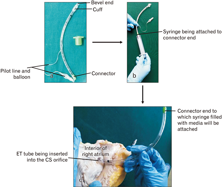
Fig. 1 Steps in the development of device. a, parts of endotracheal (ET) tube; b, syringe being attached; c, positioning of the device in the coronary sinus (CS) orifice.
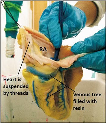
Fig. 2 The interior of right atrium (RA) is opened and device is shown in position inside the coronary sinus orifice. The coronary veins appear blue, due to the colored resin inside them.

Fig. 3 Saponified fat on the cast surface.

Fig. 4 Final cast. (A) Left oblique view of the heart cast. (B) Diaphragmatic surface of the heart cast. The veins are blue and arteries are red.
Reference
-
1. Haenssgen K, Makanya AN, Djonov V. Casting materials and their application in research and teaching. Microsc Microanal. 2014; 20:493–513.
Article2. Aharinejad SH. Corrosion casting: a historical review. In : Aharinejad SH, Lametschwandtner A, editors. Microvascular Corrosion Casting in Scanning Electron Microscopy. Vienna: Springer-Verlag;1992. p. 3–11.3. Viggiano D, Sangiorgi S, Reguzzoni M, Manelli A, Marano L, Dell'Orbo C, Passiatore C. A new method to make vascular and bronchial casts of voluminous organs. Eur J Morphol. 2003; 41:161–165.4. Meyer EP, Beer GM, Lang A, Manestar M, Krucker T, Meier S, Mihic-Probst D, Groscurth P. Polyurethane elastomer: a new material for the visualization of cadaveric blood vessels. Clin Anat. 2007; 20:448–454.
Article5. De Sordi N, Bombardi C, Chiocchetti R, Clavenzani P, Trerè C, Canova M, Grandis A. A new method of producing casts for anatomical studies. Anat Sci Int. 2014; 89:255–265.
Article6. Johnson M. Detergents: Triton X-100, Tween-20, and more [Internet]. Labome;2019. cited 2019 Jan 2. Available from: https://www.labome.com/method/Detergents-Triton-X-100-Tween-20-and-More.html.7. Sims PA, Albrecht RM. Improved tissue corrosion of vascular casts: a quantitative filtration method used to compare tissue corrosion in various concentrations of sodium and potassium hydroxide. Scanning Microsc. 1993; 7:637–642.8. Verli FD, Rossi-Schneider TR, Schneider FL, Yurgel LS, de Souza MA. Vascular corrosion casting technique steps. Scanning. 2007; 29:128–132.
Article
- Full Text Links
-
- Actions
-
Cited
- CITED
-
- Close
- Share
-
- Similar articles
-
- Modified soft tissue cast for fixed partial denture: a technique
- Comparison of the fit of cast gold crowns fabricated from the digital and the conventional impression techniques
- Role of span length in the adaptation of implant-supported cobalt chromium frameworks fabricated by three techniques
- Hydrothorax and Broken Catheters from Central Venous Catheterization
- The Treatment of Femoral Shaft Fractures by Cast Brace
