Triangles of the neck: a review with clinical/surgical applications
- Affiliations
-
- 1Seattle Science Foundation, Seattle, WA, USA. joei@seattlesciencefoundation.org
- 2Dental and Oral Medical Center, Kurume University School of Medicine, Kurume, Fukuoka, Japan.
- 3Division of Gross and Clinical Anatomy, Department of Anatomy, Kurume University School of Medicine, Kurume, Japan.
- 4Department of Anatomical Sciences, St. George's University, St. George's, Grenada, West Indies.
- KMID: 2451214
- DOI: http://doi.org/10.5115/acb.2019.52.2.120
Abstract
- The neck is a geometric region that can be studied and operated using anatomical triangles. There are many triangles of the neck, which can be useful landmarks for the surgeon. A better understanding of these triangles make surgery more efficient and avoid intraoperative complications. Herein, we provide a comprehensive review of the triangles of the neck and their clinical and surgical applications.
MeSH Terms
Figure
-
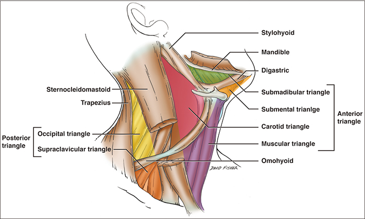
Fig. 1 Schematic drawing of the triangles and anatomical structures in the right neck.
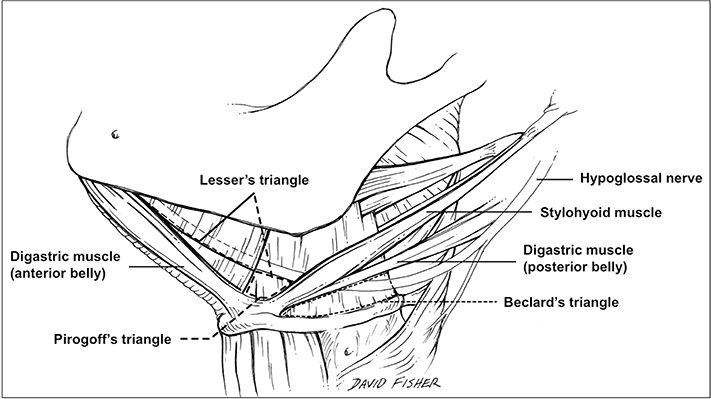
Fig. 2 Schematic drawing of Béclard's, Lesser's, and Pirogov's triangles in the left neck. Modified from Tubbs et al. Surg Radiol Anat 2011;33:53–7 [2], with permission of R. Shane Tubbs holds the copyright.
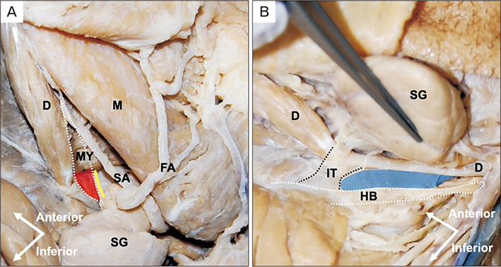
Fig. 3 Lateral view of the left neck on a formalin-fixed cadaveric dissection (region of the submandibular triangle). (A) White dotted line is Lesser's triangle, and red highlighted area is Béclard's triangle. (B) Blue highlighted area is Pirogov's triangle. D, digastric muscle; FA, facial artery; HB, hyoid bone; IT, intermediate tendon of digastric muscle; M, mandible; MY, mylohyoid muscle; SA, submental artery; SG, submandibular gland.
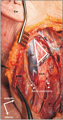
Fig. 4 Farabeuf's triangle of the right lateral neck on a fresh-frozen cadaver dissection (white solid line). CCA, common carotid artery; CFV, common facial vein; D, digastric muscle; HN, hypoglossal nerve; IJV, internal jugular vein; RMV, retromandibular vein; SCM, sternocleidomastoid muscle; ST, sternothyroid muscle.
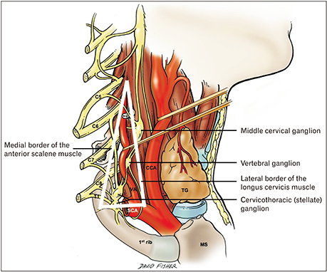
Fig. 5 Schematic drawing of the triangle of the vertebral artery on the right lateral neck region (white solid line). CCA, common carotid artery; MS, manubrium of the sternum; SCA, subclavian artery; TG, thyroid gland; VA, vertebral artery.
Cited by 2 articles
-
Localizing the nerve to the mylohyoid using the mylohyoid triangle
Joe Iwanaga, Hee-Jin Kim, Grzegorz Wysiadecki, Kyoichi Obata, Yosuke Harazono, Soichiro Ibaragi, R. Shane Tubbs
Anat Cell Biol. 2021;54(3):304-307. doi: 10.5115/acb.21.019.An anatomical investigation of the suboccipital- and inferior suboccipital triangles
Kirsten Shannon Regan, Gerda Venter
Anat Cell Biol. 2023;56(3):350-359. doi: 10.5115/acb.23.015.
Reference
-
1. Tubbs RS, Rasmussen M, Loukas M, Shoja M, Mortazavi M, Cohen-Gadol AA. Use of the triangle of farabeuf for neurovascular procedures of the neck. Biomed Int. 2011; 2:39–42.2. Tubbs RS, Rasmussen M, Loukas M, Shoja MM, Cohen-Gadol AA. Three nearly forgotten anatomical triangles of the neck: triangles of Beclard, Lesser and Pirogoff and their potential applications in surgical dissection of the neck. Surg Radiol Anat. 2011; 33:53–57.
Article3. Hiatt JL. Textbook of head and neck anatomy. Philadelphia, PA: Lippincott Williams and Wilkins;2001.4. Davies JC, Ravichandiran M, Agur AM, Fattah A. Evaluation of clinically relevant landmarks of the marginal mandibular branch of the facial nerve: a three-dimensional study with application to avoiding facial nerve palsy. Clin Anat. 2016; 29:151–156.
Article5. Jamieson GG. Anatomy of general surgical operations. Edinburgh: Elsevier;2006.6. Deaver JB. Surgical anatomy of the human body. Philadelphia, PA: Blakiston's Sons & Co.;1926.7. van Es RJ, Thuau H. Pirogoff's triangle revisited: an alternative site for microvascular anastomosis to the lingual artery: a technical note. Int J Oral Maxillofac Surg. 2000; 29:207–209.
Article8. Burke RH, Masch GL. Lingual artery hemorrhage. Oral Surg Oral Med Oral Pathol. 1986; 62:258–261.
Article9. He P, Truong MK, Adeeb N, Tubbs RS, Iwanaga J. Clinical anatomy and surgical significance of the lingual foramina and their canals. Clin Anat. 2017; 30:194–204.
Article10. Pirogoff NI. Anatome topographica sectionibus per corpus humanum congelatum triplice directione ductis illustrata. Petersburg: Fleischer;1852.11. Skandalakis JE, Gray SW, Ricketts RR, Richardson DD. Skandalakis' surgical anatomy: the embryologic and anatomic basis of modern surgery. Athens: Paschalidis Medical Publications;2004.12. Homze EJ, Harn SD, Bavitz BJ. Extraoral ligation of the lingual artery: an anatomic study. Oral Surg Oral Med Oral Pathol Oral Radiol Endod. 1997; 83:321–324.13. Béclard PA, Knox R. Elements of general anatomy. Edinburgh: MacLachlan and Stewart;1830.14. Henry AK. Extensile exposure. Edinburgh: Churchill Livingstone;1973.15. Campbell WF. A text-book of surgical anatomy. Philadelphia, PA: WB Saunders Company;1922.16. Standring S. Gray's anatomy: the anatomical basis of clinical practice. 41st ed. London: Elsevier Health Sciences;2015.17. Bergenfelz A, Jansson S, Kristoffersson A, Mårtensson H, Reihnér E, Wallin G, Lausen I. Complications to thyroid surgery: results as reported in a database from a multicenter audit comprising 3,660 patients. Langenbecks Arch Surg. 2008; 393:667–673.
Article18. Cheung NH, Napolitano LM. Tracheostomy: epidemiology, indications, timing, technique, and outcomes. Respir Care. 2014; 59:895–915.
Article19. Laskin DM. Anatomic considerations in diagnosis and treatment of odontogenic infections. J Am Dent Assoc. 1964; 69:308–316.20. Amar AP, Heck CN, Levy ML, Smith T, DeGiorgio CM, Oviedo S, Apuzzo ML. An institutional experience with cervical vagus nerve trunk stimulation for medically refractory epilepsy: rationale, technique, and outcome. Neurosurgery. 1998; 43:1265–1276.
Article21. Tubbs RS, Loukas M, Shoja MM, Salter EG, Oakes WJ, Blount JP. Approach to the cervical portion of the vagus nerve via the posterior cervical triangle: a cadaveric feasibility study with potential use in vagus nerve stimulation procedures. J Neurosurg Spine. 2006; 5:540–542.
Article22. Tubbs RS, Salter EG, Oakes WJ. Anatomic landmarks for nerves of the neck: a vade mecum for neurosurgeons. Neurosurgery. 2005; 56:256–260.
Article23. Tubbs RS, Salter EG, Wellons JC 3rd, Blount JP, Oakes WJ. Superficial landmarks for the spinal accessory nerve within the posterior cervical triangle. J Neurosurg Spine. 2005; 3:375–378.
Article24. Cesmebasi A, Spinner RJ. An anatomic-based approach to the iatrogenic spinal accessory nerve injury in the posterior cervical triangle: How to avoid and treat it. Clin Anat. 2015; 28:761–766.
Article25. Graves MJ, Henry BM, Hsieh WC, Sanna B, PĘkala PA, Iwanaga J, Loukas M, Tomaszewski KA. Origin and prevalence of the accessory phrenic nerve: a meta-analysis and clinical appraisal. Clin Anat. 2017; 30:1077–1082.
Article26. Talbot RW. Anatomical pitfall of subclavian venepuncture. Ann R Coll Surg Engl. 1978; 60:317–319.27. Prates Júnior AG, Vasques LC, Bordoni LS. Anatomical variations of the phrenic nerve: an actualized review. J Morphol Sci. 2015; 32:53–56.
Article28. Loukas M, Kinsella CR Jr, Louis RG Jr, Gandhi S, Curry B. Surgical anatomy of the accessory phrenic nerve. Ann Thorac Surg. 2006; 82:1870–1875.
Article29. Sharma MS, Loukas M, Spinner RJ. Accessory phrenic nerve: a rarely discussed common variation with clinical implications. Clin Anat. 2011; 24:671–673.
Article30. Bielamowicz SA, Storper IS, Jabour BA, Lufkin RB, Hanafee WN. Spaces and triangles of the head and neck. Head Neck. 1994; 16:383–388.
Article31. Virchow R. Zur diagnose der krebse im unterleibe. Med Reform. 1848; 45:248.32. Troisier E. Les ganglions sus-claviculaires dans le cancer de l'estomac. Bull Mem Soc Med Hop Paris. 1886; 3:394–398.33. Morgenstern L. The Virchow-Troisier node: a historical note. Am J Surg. 1979; 138:703.
Article34. Wechsler RJ, Rao VM, Newman LM. The subclavian triangle: CT analysis. AJR Am J Roentgenol. 1989; 152:313–317.
Article35. Hollinishead WH. Anatomy for surgeons. Vol. 2. The thorax, abdomen and pelvis. New York: Harper and Row;1969.36. Loukas M, Tubbs RS. An accessory muscle within the suboccipital triangle. Clin Anat. 2007; 20:962–963.
Article37. Tubbs RS, Salter EG, Wellons JC, Blount JP, Oakes WJ. Landmarks for the identification of the cutaneous nerves of the occiput and nuchal regions. Clin Anat. 2007; 20:235–238.
Article38. Tubbs RS, Salter EG, Wellons JC 3rd, Blount JP, Oakes WJ. The triangle of the vertebral artery. Neurosurgery. 2005; 56:2 Suppl. 252–255.
Article39. Schaeffer JP. Morris' human anatomy: a complete systematic treatise. 11th ed. New York: Blakiston;1953.40. Gardner E, Gray DJ, O'Rahilly R. Anatomy: a regional study of human structure. 4th ed. Philadelphia, PA: W.B. Saunders Co.;1975.41. Lang J. Arteries of the neck in clinical anatomy of the cervical spine. New York: Thieme;1993.42. Shen XH, Xue HD, Chen Y, Wang M, Mirjalili SA, Zhang ZH, Ma C. A reassessment of cervical surface anatomy via CT scan in an adult population. Clin Anat. 2017; 30:330–335.
Article43. Badshah M, Soames R, Ibrahim M, Khan MJ, Khan A. Surface anatomy of major anatomical landmarks of the neck in an adult population: a Ct evaluation of vertebral level. Clin Anat. 2017; 30:781–787.44. Youmans J. Neurological surgery: a comprehensive reference guide to the diagnosis and management of neurosurgical problems. 2nd ed. Philadelphia, PA: W.B. Saunders Co.;1982.45. Rusnak-Smith S, Moffat M, Rosen E. Anatomical variations of the scalene triangle: dissection of 10 cadavers. J Orthop Sports Phys Ther. 2001; 31:70–80.
Article46. Harry WG, Bennett JD, Guha SC. Scalene muscles and the brachial plexus: anatomical variations and their clinical significance. Clin Anat. 1997; 10:250–252.
Article47. Klaassen Z, Sorenson E, Tubbs RS, Arya R, Meloy P, Shah R, Shirk S, Loukas M. Thoracic outlet syndrome: a neurological and vascular disorder. Clin Anat. 2014; 27:724–732.
Article48. Atasoy E. Thoracic outlet syndrome: anatomy. Hand Clin. 2004; 20:7–14.
Article
- Full Text Links
-
- Actions
-
Cited
- CITED
-
- Close
- Share
-
- Similar articles
-
- Ten Triangles around Cavernous Sinus for Surgical Approach, Described by Schematic Diagram and Three Dimensional Models with the Sectioned Images
- An anatomical investigation of the suboccipitaland inferior suboccipital triangles
- Ansa cervicalis: a comprehensive review of its anatomy, variations, pathology, and surgical applications
- The applications of endoscopic surgery in department of otorhinolaryngology-head and neck surgery
- Computational Simulation of Multiple Cannulated Screw Fixation for Femoral Neck Fractures and the Anatomic Features for Clinical Applications
