Brachial-Ankle Pulse Wave Velocity as a Screen for Arterial Stiffness: A Comparison with Cardiac Magnetic Resonance
- Affiliations
-
- 1Division of Cardiology, Department of Medicine, Heart Vascular Stroke Institute, Samsung Medical Center, Sungkyunkwan University School of Medicine, Seoul, Korea. dkkim@skku.edu
- 2Department of Radiology, Heart Vascular Stroke Institute, Samsung Medical Center, Sungkyunkwan University School of Medicine, Seoul, Korea.
- KMID: 2450333
- DOI: http://doi.org/10.3349/ymj.2015.56.3.617
Abstract
- PURPOSE
Despite technical simplicity and the low cost of brachial-ankle pulse wave velocity (BA-PWV), its use has been hampered by a lack of data supporting its usefulness and reliability. The aim of this study was to evaluate the usefulness of BA-PWV to measure aortic stiffness in comparison to using cardiovascular magnetic resonance (CMR).
MATERIALS AND METHODS
A total of 124 participants without cardiovascular risk factors volunteered for this study. BA-PWV was measured using a vascular testing device. On the same day, using CMR, cross-sectional areas for distensibility and average blood flow were measured at four aortic levels: the ascending, upper thoracic descending, lower thoracic descending, and abdominal aorta.
RESULTS
Compared to PWV measured by CMR, BA-PWV values were significantly higher and the differences therein were similar in all age groups (all p<0.001). There was a significant correlation between BA-PWV and PWV by CMR (r=0.697, p<0.001). Both BA-PWV and PWV by CMR were significantly and positively associated with age (r=0.652 and 0.724, p<0.001). The reciprocal of aortic distensibility also demonstrated a statistically significant positive correlation with BA-PWV (r=0.583 to 0.673, all p<0.001).
CONCLUSION
BA-PWV was well correlated with central aortic PWV and distensibility, as measured by CMR, regardless of age and sex.
Keyword
MeSH Terms
Figure
-
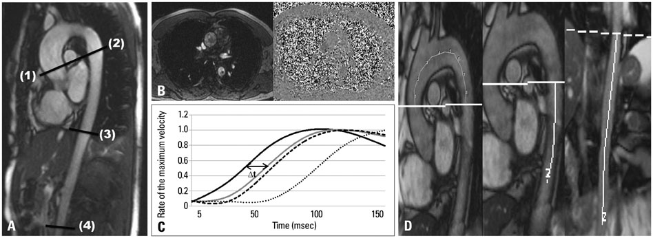
Fig. 1 (A) Contrast enhanced MR imaging showing the aorta in the long axis. Each level perpendicular to the aorta was applied for gradient echo imaging with velocity encoding. Distensibility was assessed at these four levels: ascending (level 1), upper descending thoracic (level 2), lower descending thoracic (level 3), and abdominal aorta (level 4). PWV was calculated as the ratio of the distance between two levels and the time difference between arrivals of flow waves at these levels. Consequently, the PWV-CMR value was determined in three regions of the aorta: proximal (level 1-2), descending thoracic (level 2-3), and abdominal aorta (level 3-4). (B, C, and D) Aortic PWV assessment using CMR. (B) Velocity encoded MR images of the ascending aorta. Flow curves of 4 regions-ascending aorta, upper descending thoracic aorta, lower descending thoracic aorta, and abdominal aorta, respectively. (C) This graph shows the transit delay (Δt) of the systolic flow curves in the upper descending thoracic aorta relative to the ascending aorta. The transit time (Δt) is determined from the midpoints of the systolic up-slope at 40-60% of the maximum velocity in the waveform in the flow versus time curves. (D) Sagittal CMR image that shows the distance between each aortic level is measured along the midline through the aorta. Aortic PWV was obtained by distance/Δt. PWV, pulse wave velocity; CMR, cardiovascular magnetic resonance.
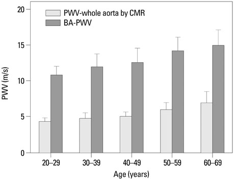
Fig. 2 Comparison of the BA-PWV and aortic PWV by cardiac magnetic resonance imaging according to age group. Each bar is expressed as the mean and standard deviation. PWV, pulse wave velocity; BA-PWV, brachial-ankle PWV; CMR, cardiovascular magnetic resonance.
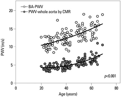
Fig. 3 Relationship between age and BA-PWV and aortic PWV obtained using cardiac magnetic resonance imaging. PWV, pulse wave velocity; BA-PWV, brachial-ankle PWV; CMR, cardiovascular magnetic resonance.

Fig. 4 (A) Relationship between the BA-PWV and aortic PWV by cardiac magnetic resonance imaging and corresponding Bland-Altman plots. (B) Relationship between the CF-PWV and aortic PWV by cardiac magnetic resonance imaging and corresponding Bland-Altman plots. PWV, pulse wave velocity; BA-PWV, brachial-ankle PWV; CF-PWV, carotid-femoral PWV; CMR, cardiovascular magnetic resonance.
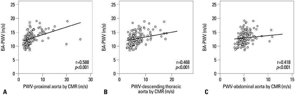
Fig. 5 Relationship between the BA-PWV and the regional aortic PWV according to the measured sites: proximal aorta (A), descending thoracic aorta (B), and abdominal aorta (C). PWV, pulse wave velocity; BA-PWV, brachial-ankle PWV; CMR, cardiovascular magnetic resonance.
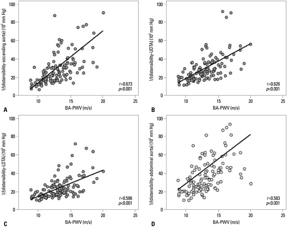
Fig. 6 Relationship between the BA-PWV and the regional aortic distensibility at the measured sites: ascending aorta (A), upper descending thoracic aorta (B), lower descending thoracic aorta (C), and abdominal aorta (D). BA-PWV, brachial-ankle PWV; LDTA, lower descending thoracic aorta; UDTA, upper descending thoracic aorta.
Reference
-
1. Sutton-Tyrrell K, Najjar SS, Boudreau RM, Venkitachalam L, Kupelian V, Simonsick EM, et al. Elevated aortic pulse wave velocity, a marker of arterial stiffness, predicts cardiovascular events in well-functioning older adults. Circulation. 2005; 111:3384–3390.
Article2. Choi CU, Park EB, Suh SY, Kim JW, Kim EJ, Rha SW, et al. Impact of aortic stiffness on cardiovascular disease in patients with chest pain: assessment with direct intra-arterial measurement. Am J Hypertens. 2007; 20:1163–1169.3. Laurent S, Boutouyrie P, Asmar R, Gautier I, Laloux B, Guize L, et al. Aortic stiffness is an independent predictor of all-cause and cardiovascular mortality in hypertensive patients. Hypertension. 2001; 37:1236–1241.
Article4. Laurent S, Cockcroft J, Van Bortel L, Boutouyrie P, Giannattasio C, Hayoz D, et al. Expert consensus document on arterial stiffness: methodological issues and clinical applications. Eur Heart J. 2006; 27:2588–2605.
Article5. Klocke R, Cockcroft JR, Taylor GJ, Hall IR, Blake DR. Arterial stiffness and central blood pressure, as determined by pulse wave analysis, in rheumatoid arthritis. Ann Rheum Dis. 2003; 62:414–418.
Article6. Boutouyrie P, Tropeano AI, Asmar R, Gautier I, Benetos A, Lacolley P, et al. Aortic stiffness is an independent predictor of primary coronary events in hypertensive patients: a longitudinal study. Hypertension. 2002; 39:10–15.
Article7. Shiotani A, Motoyama M, Matsuda T, Miyanishi T. Brachial-ankle pulse wave velocity in Japanese university students. Intern Med. 2005; 44:696–701.
Article8. Aso K, Miyata M, Kubo T, Hashiguchi H, Fukudome M, Fukushige E, et al. Brachial-ankle pulse wave velocity is useful for evaluation of complications in type 2 diabetic patients. Hypertens Res. 2003; 26:807–813.
Article9. Yamashina A, Tomiyama H, Takeda K, Tsuda H, Arai T, Hirose K, et al. Validity, reproducibility, and clinical significance of noninvasive brachial-ankle pulse wave velocity measurement. Hypertens Res. 2002; 25:359–364.
Article10. Westenberg JJ, de Roos A, Grotenhuis HB, Steendijk P, Hendriksen D, van den Boogaard PJ, et al. Improved aortic pulse wave velocity assessment from multislice two-directional in-plane velocity-encoded magnetic resonance imaging. J Magn Reson Imaging. 2010; 32:1086–1094.
Article11. Ibrahim el-SH, Johnson KR, Miller AB, Shaffer JM, White RD. Measuring aortic pulse wave velocity using high-field cardiovascular magnetic resonance: comparison of techniques. J Cardiovasc Magn Reson. 2010; 12:26.
Article12. Giannattasio C, Cesana F, Maestroni S, Salvioni A, Maloberti A, Nava S, et al. Comparison of echotracking and magnetic resonance assessment of abdominal aorta distensibility and relationships with pulse wave velocity. Ultrasound Med Biol. 2011; 37:1970–1976.
Article13. Sugawara J, Hayashi K, Yokoi T, Cortez-Cooper MY, DeVan AE, Anton MA, et al. Brachial-ankle pulse wave velocity: an index of central arterial stiffness? J Hum Hypertens. 2005; 19:401–406.
Article14. Cortez-Cooper MY, Supak JA, Tanaka H. A new device for automatic measurements of arterial stiffness and ankle-brachial index. Am J Cardiol. 2003; 91:1519–1522.
Article15. Voges I, Jerosch-Herold M, Hedderich J, Pardun E, Hart C, Gabbert DD, et al. Normal values of aortic dimensions, distensibility, and pulse wave velocity in children and young adults: a cross-sectional study. J Cardiovasc Magn Reson. 2012; 14:77.
Article16. Van Bortel LM, Laurent S, Boutouyrie P, Chowienczyk P, Cruickshank JK, De Backer T, et al. Expert consensus document on the measurement of aortic stiffness in daily practice using carotid-femoral pulse wave velocity. J Hypertens. 2012; 30:445–448.
Article17. Tanaka H, Munakata M, Kawano Y, Ohishi M, Shoji T, Sugawara J, et al. Comparison between carotid-femoral and brachial-ankle pulse wave velocity as measures of arterial stiffness. J Hypertens. 2009; 27:2022–2027.
Article18. Benetos A, Laurent S, Hoeks AP, Boutouyrie PH, Safar ME. Arterial alterations with aging and high blood pressure. A noninvasive study of carotid and femoral arteries. Arterioscler Thromb. 1993; 13:90–97.
Article19. van der Heijden-Spek JJ, Staessen JA, Fagard RH, Hoeks AP, Boudier HA, van Bortel LM. Effect of age on brachial artery wall properties differs from the aorta and is gender dependent: a population study. Hypertension. 2000; 35:637–642.
Article20. Cameron JD, Bulpitt CJ, Pinto ES, Rajkumar C. The aging of elastic and muscular arteries: a comparison of diabetic and nondiabetic subjects. Diabetes Care. 2003; 26:2133–2138.21. Munakata M, Ito N, Nunokawa T, Yoshinaga K. Utility of automated brachial ankle pulse wave velocity measurements in hypertensive patients. Am J Hypertens. 2003; 16:653–657.
Article
- Full Text Links
-
- Actions
-
Cited
- CITED
-
- Close
- Share
-
- Similar articles
-
- Increased arterial stiffness causing resistant hypertension in an adolescent with Neurofibromatosis type 1
- Uric Acid Level Has a J-Shaped Association with Arterial Stiffness in Korean Postmenopausal Women
- Pulse wave velocity and ankle brachial index in normal adolescents
- Correlation of Arterial Stiffness and Bone Mineral Density by Measuring Brachial-Ankle Pulse Wave Velocity in Healthy Korean Women
- Usefulness of Brachial-Ankle Pulse Wave Velocity and Ankle-Brachial Index as Predictors of Early Age-Related Maculopathy
