Imaging Sci Dent.
2018 Dec;48(4):255-259. 10.5624/isd.2018.48.4.255.
Development of a new ball-type phantom for evaluation of the image layer of panoramic radiography
- Affiliations
-
- 1Department of Oral and Maxillofacial Radiology and Dental Research Institute, School of Dentistry, Seoul National University, Seoul, Korea. raylee@snu.ac.kr
- KMID: 2450165
- DOI: http://doi.org/10.5624/isd.2018.48.4.255
Abstract
- PURPOSE
This study proposes a new ball-type phantom for evaluation of the image layer of panoramic radiography.
MATERIALS AND METHODS
The arch shape of an acrylic resin phantom was derived from average data on the lower dental arch in Korean adult males. Metal balls with a 2-mm diameter were placed along the center line of the phantom at a 4-mm mesiodistal interval. Additional metal balls were placed along the 22 arch-shaped lines that ran parallel to the center line at 2-mm buccolingual intervals. The height of each ball in the horizontal plane was spaced by 2.5 mm, and consequently, the balls appeared oblique when viewed from the side. The resulting phantom was named the Panorama phantom. The distortion rate of the balls in the acquired image was measured by automatically calculating the difference between the vertical and horizontal length using MATLAB®. Image layer boundaries were obtained by applying various distortion rate thresholds.
RESULTS
Most areas containing metal balls (91.5%) were included in the image layer with a 50% distortion rate threshold. When a 5% distortion rate threshold was applied, the image layer was formed with a small buccolingual width along the arch-shaped center line. However, it was medially located in the temporomandibular joint region.
CONCLUSION
The Panorama phantom could be used to evaluate the image layer of panoramic radiography, including all mesiodistal areas with large buccolingual width.
Keyword
MeSH Terms
Figure
-
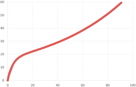
Fig. 1 Graph of the adjusted formula. The continuous line obtained from the graph is defined as the center line of the phantom.

Fig. 2 A. Final design of the Panorama phantom. The balls appeared oblique when viewed from the side. B. Actual model of the Panorama phantom. It was made of acrylic resin and metal balls with a diameter of 2 mm.
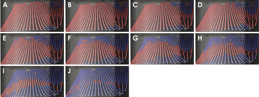
Fig. 3 Distortion rate thresholds of the obtained images of metal balls. The balls that satisfied the distortion rate threshold are displayed in red. (A–J) Several image layer boundaries were obtained by applying various distortion rate thresholds, from 50% to 5% with 5% decrements.
Reference
-
1. Hassen SM, Manson-Hing LR. A study of the zone of sharpness of three panoramic x-ray machines and the effect of screen speed on the sharpness zone. Oral Surg Oral Med Oral Pathol. 1982; 54:242–249.
Article2. Paiboon C, Manson-Hing LR. Effect of border sharpness on the size and position of the focal trough of panoramic x-ray machines. Oral Surg Oral Med Oral Pathol. 1985; 60:670–676.
Article3. Rondon RH, Pereira YC, do Nascimento GC. Common positioning errors in panoramic radiography: a review. Imaging Sci Dent. 2014; 44:1–6.
Article4. Patel JR. The focal trough of the Autopan panoramic dental x-ray machine. Oral Surg Oral Med Oral Pathol. 1989; 68:115–118.
Article5. Brown CE Jr, Christen AC, Jerman AC. Dimensions of the focal trough in panoramic radiography. J Am Dent Assoc. 1972; 84:843–847.
Article6. Lund TM, Manson-Hing LR. A study of the focal troughs of three panoramic dental x-ray machines. Part I. The area of sharpness. Oral Surg Oral Med Oral Pathol. 1975; 39:318–328.7. Nam JH, Lee KS. A study of mandibular dental arch form of the Korean with normal occlusion. Korean J Orthod. 1996; 26:535–546.8. Lee SJ, Moon SC, Kim TW, Nahm DS, Chang YI. Tooth size and arch parameters of normal occlusion in a large Korean sample. Korean J Orthod. 2004; 34:473–480.9. Kim KH, Choy KC, Kim HG, Park KH. Cephalometric norms of the hard tissues of Korean for orthognathic surgery. J Korean Assoc Oral Maxillofac Surg. 2001; 27:221–230.10. Ahn HK. Observation of condyles by roentgenographic cephalogram. J Korean Acad Oral Maxillofac Radiol. 1984; 14:135–140.11. Dibbets JM, Nolte K. Effect of magnification on lateral cephalometric studies. Am J Orthod Dentofacial Orthop. 2002; 122:196–201.
Article
- Full Text Links
-
- Actions
-
Cited
- CITED
-
- Close
- Share
-
- Similar articles
-
- A study of panoramic focal trough for the six-year-old child
- Erratum to: Development of a new ball-type phantom for evaluation of the image layer of panoramic radiography
- Quantitative localization of impacted mesiodens using panoramic and periapical radiographs
- Reference line-pair values of panoramic radiographs using an arch-form phantom stand to assess clinical image quality
- Effective dose from direct and indirect digital panoramic units
