Inhibition of Indoleamine 2,3-Dioxygenase Enhances the Therapeutic Efficacy of Immunogenic Chemotherapeutics in Breast Cancer
- Affiliations
-
- 1Department of Clinical Medicine, Clinical Medical College of Shandong University, Jinan, China.
- 2Department of General Surgery, the First Affiliated Hospital of University of Science and Technology of China, Division of Life Sciences and Medicine, University of Science and Technology of China, Hefei, China.
- 3Department of Hepatic Surgery, the First Affiliated Hospital of University of Science and Technology of China, Division of Life Sciences and Medicine, University of Science and Technology of China, Hefei, China. wdjia2018@sina.com
- KMID: 2450115
- DOI: http://doi.org/10.4048/jbc.2019.22.e23
Abstract
- PURPOSE
Breast cancer has become a major public health threat in the current society. Anthracycline doxorubicin (DOX) is a widely used drug in breast cancer chemotherapy. We aimed to investigate the immunogenic death of breast tumor cells caused by DOX, and detect the effects of combination of DOX and a small molecule inhibitor in tumor engrafted mouse model.
METHODS
We used 4T1 breast cancer cells to examine the anthracycline DOX-mediated immunogenic death of breast tumor cells by assessing the calreticulin exposure and adenosine triphosphate and high mobility group box 1 release. Using 4T1 tumor cell-engrafted mouse model, we also detected the expression of indoleamine 2,3-dioxygenase (IDO) in tumor tissues after DOX treatment and further explored whether the specific small molecule IDO1 inhibitor NLG919 combined with DOX, can exhibit better therapeutic effects on breast cancer.
RESULTS
DOX induced immunogenic cell death of murine breast cancer cells 4T1 as well as the upregulation of IDO1. We also found that treatment with NLG919 enhanced kynurenine inhibition in a dose-dependent manner. IDO1 inhibition reversed CD8+ T cell suppression mediated by IDO-expressing 4T1 murine breast cancer cells. Compared to the single agent or control, combination of DOX and NLG919 significantly inhibited the tumor growth, indicating that the 2 drugs exhibit synergistic effect. The combination therapy also increased the expression of transforming growth factor-β, while lowering the expressions of interleukin-12p70 and interferon-γ.
CONCLUSION
Compared to single agent therapy, combination of NLG919 with DOX demonstrated better therapeutic effects in 4T1 murine breast tumor model. IDO inhibition by NLG919 enhanced the therapeutic efficacy of DOX in breast cancer, achieving synergistic effect.
MeSH Terms
Figure
-

Figure 1 DOX induces immunogenic cell death of murine breast cancer cells 4T1. (A) The surface exposure of CRT was determined by flow cytometry among viable (propidium iodine-negative) cells after treatment with different concentrations of DOX for 24 hours. DOX-treated cells were stained with propidium iodine and FITC-labeled anti-CRT antibodies according to the manufacturer's instructions. (B) The percentage of CRT-positive cells in PI-negative cells was quantified based on based on the results of flow cytometry. (C) S-HMGB1 of 4T1 cells treated with different concentrations of DOX was measured by western blotting, and BSA was used as the loading control. (D) Level of released ATP was determined by a chemiluminescent ATP Determination Kit. (E) Animal vaccination, using 2 rounds of s.c. injection of DOX-treated dying 4T1 cells at 7 days apart, followed by s.c. injection of live cells on the contralateral side. Two weeks later, the number of mice without tumor on contralateral side was counted. Successful tumor growth inhibition at the challenge site is suggestive of immune interference. (F) The number of mice without visible tumor on contralateral side 2 weeks post second injection. Data represent means ± standard deviation. DOX = doxorubicin; ATP = adenosine triphosphate; BSA = bovine serum albumin; DOX = doxorubicin; CRT = calreticulin; FITC = fluorescein isothiocyanate; PI = propidium iodide; SD = standard deviation; S-HMGB1 = supernatant high mobility group box 1; s.c. = subcutaneous. *p < 0.05; †p < 0.01; ‡p < 0.001 (versus control group).
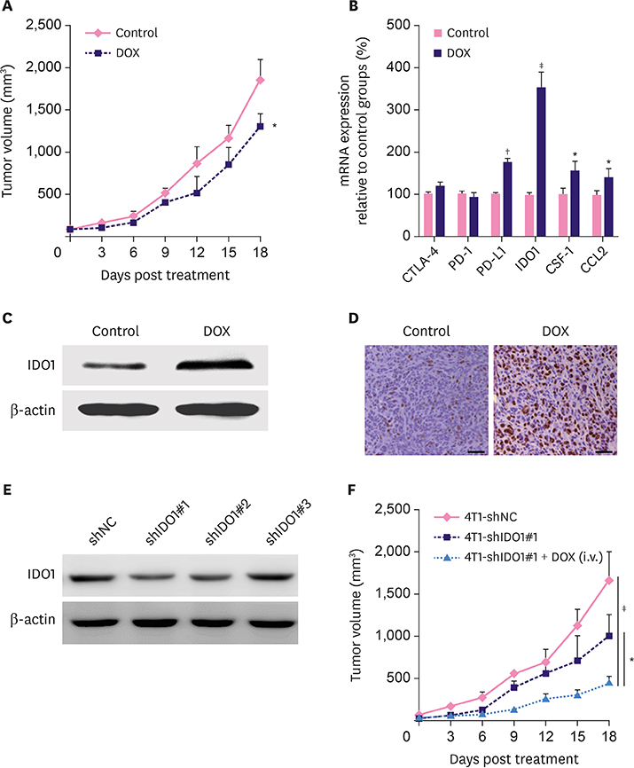
Figure 2 DOX treatment induces upregulation of IDO1 and IDO1 impairs the therapeutic efficacy of DOX. (A) Slight inhibition of tumor growth by DOX in 4T1 tumor-bearing BALB/c mice (n = 6). DOX was i.v. injected at the dosage of 5.0 mg/kg for 5 times after every 3 days. (B) The mRNA expressions of CTLA-4, PD-1, PD-L1, IDO1, CSF-1, and CCL2 examined by real time polymerase chain reaction. The mRNA levels of these genes were normalized against the expression level of the housekeeping gene GAPDH. The IDO1 protein expression in tumor tissues from mice treated with phosphate buffer saline solution and DOX were examined by western blotting (C) and immunohistochemistry (D). (E) Protein levels of IDO1 in 4T1-shNC and 4T1-shIDO1 cells detected by western blot. β-actin was used as loading control. (F) Tumor growth in 4T1-shNC and 4T1-shIDO1 tumor bearing BALB/c mice. 4T1-shIDO1 tumor bearing BALB/c mice were injected with DOX at the dosage of 5.0 mg/kg. Data represent means ± standard deviation. DOX = doxorubicin; mRNA = messenger RNA; CTLA-4 = cytotoxic T-lymphocyte-associated protein 4; PD-1 = programmed cell death 1; PD-L1 = programmed cell death 1; CSF-1 = cerebrospinal fluid 1; CCL2 = chemokine (C-C motif) ligand 2; IDO1 = indoleamine 2,3-dioxygenase 1; shNC = short hairpin RNA non-target control; shIDO1 = short hairpin RNAs targeting indoleamine 2,3-dioxygenase 1. *p < 0.05; †p < 0.01; ‡p < 0.001.
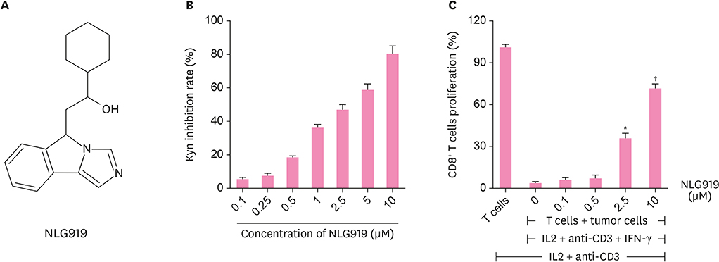
Figure 3 In vitro activities of IDO1-specific inhibitor (NLG919). (A) Molecular structural formula of NLG919, a highly selective inhibitor of IDO1. (B) NLG919 inhibited IDO1 enzyme activity in vitro. The 4T1 cells were treated with IFN-γ together with different concentrations of NLG919. Kyn level in supernatant was measured 2 days later using high performance liquid chromatography mass spectrometry. The 4T1 cells treated only with IFN-γ were set as blank control. (C) IDO1 inhibition reversed T-cell suppression mediated by IDO-expressing murine breast cancer cells (4T1). The 4T1 cells and splenocytes were co-cultured, and then, treated with IL-2, anti-CD3 antibody, and IFN-γ together with NLG919 for 3 days. The proliferation of CD8+ T cells was examined by fluorescence-activated cell sorting analysis. Data represent means ± standard deviation. IDO1 = indoleamine 2,3-dioxygenase 1; Kyn = kynurenine; IL = interleukin; IFN = interferon. *p < 0.05; †p < 0.01 (versus control).
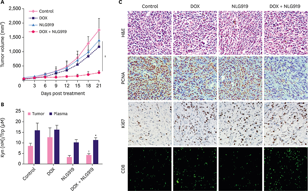
Figure 4 In vivo anti-tumor effect of DOX, NLG919, and combination of both agents. (A) Inhibition of tumor growth by various treatments in 4T1 tumor-bearing BALB/c mice (n = 6). BALB/c mice bearing 4T1 tumors of ~50 mm3 volume were treated with either phosphate buffer saline, DOX, NLG919, or a combination of DOX and NLG919 for 5 times after 3 days each; the administration dosage of DOX and NLG919 were 5.0 mg/kg (i.v.) and 20 mg/kg (orally), respectively. (B) NLG919 treatment decreased Kyn levels in tumors and plasma. Kyn/Trp ratios in tumors and plasma were determined by high performance liquid chromatography mass spectrometry at the end of the treatment. (C) H&E, PCNA, and Ki67 staining, and CD8+ T cells analyses of tumor tissues after treatment with various therapeutic agents. The PCNA-positive proliferating cells and Ki67-positive apoptotic cells are stained brown. CD8+ T cells are stained green. Data represent means ± standard deviation. DOX = doxorubicin; Kyn = kynurenine; Trp = tryptophan; PCNA = proliferating cell nuclear antigen; H&E = hematoxylin and eosin stain. *p < 0.05; †p < 0.01; ‡p < 0.001 (versus DOX group).

Figure 5 Flow cytometry analysis of immune cell subsets and ELISA analysis of cytokine expression in tumor tissues. (A) Flow cytometry gating and histogram analysis of mature DCs in the tumor tissues at the end of treatment. The mature DCs were denoted as CD80+CD86+ populations (gate in CD45+CD11b+CD11c+ cell population). (B) Flow cytometry gating and histogram analysis of cytotoxic T cells (CD8+ T cells) in the CD45+ TILs in tumor tissues from mice receiving indicated treatment. (C-E) ELISA results of cytokine production in the tumors from mice receiving indicated treatments. Data represent means ± standard deviation. ELISA = enzyme-linked immunosorbent assay; DOX = doxorubicin; DC = dendritic cell; TIL = tumor infiltrating lymphocyte; TGF = transforming growth factor; IL = interleukin; IFN = interferon. *p < 0.05; †p < 0.01 (versus DOX group).
Reference
-
1. Hutchinson L. Breast cancer: challenges, controversies, breakthroughs. Nat Rev Clin Oncol. 2010; 7:669–670.2. Siegel RL, Miller KD, Jemal A. Cancer Statistics, 2017. CA Cancer J Clin. 2017; 67:7–30.
Article3. Anampa J, Makower D, Sparano JA. Progress in adjuvant chemotherapy for breast cancer: an overview. BMC Med. 2015; 13:195.
Article4. Sledge GW, Mamounas EP, Hortobagyi GN, Burstein HJ, Goodwin PJ, Wolff AC. Past, present, and future challenges in breast cancer treatment. J Clin Oncol. 2014; 32:1979–1986.
Article5. Garg AD, More S, Rufo N, Mece O, Sassano ML, Agostinis P, et al. Trial watch: Immunogenic cell death induction by anticancer chemotherapeutics. OncoImmunology. 2017; 6:e1386829.
Article6. Kroemer G, Galluzzi L, Kepp O, Zitvogel L. Immunogenic cell death in cancer therapy. Annu Rev Immunol. 2013; 31:51–72.
Article7. Binnewies M, Roberts EW, Kersten K, Chan V, Fearon DF, Merad M, et al. Understanding the tumor immune microenvironment (TIME) for effective therapy. Nat Med. 2018; 24:541–550.
Article8. Lu J, Liu X, Liao YP, Salazar F, Sun B, Jiang W, et al. Nano-enabled pancreas cancer immunotherapy using immunogenic cell death and reversing immunosuppression. Nat Commun. 2017; 8:1811.
Article9. Moon YW, Hajjar J, Hwu P, Naing A. Targeting the indoleamine 2,3-dioxygenase pathway in cancer. J Immunother Cancer. 2015; 3:51.
Article10. Chen X, Parelkar SS, Henchey E, Schneider S, Emrick T. PolyMPC-doxorubicin prodrugs. Bioconjug Chem. 2012; 23:1753–1763.
Article11. Yue EW, Douty B, Wayland B, Bower M, Liu X, Leffet L, et al. Discovery of potent competitive inhibitors of indoleamine 2,3-dioxygenase with in vivo pharmacodynamic activity and efficacy in a mouse melanoma model. J Med Chem. 2009; 52:7364–7367.
Article12. Munn DH, Shafizadeh E, Attwood JT, Bondarev I, Pashine A, Mellor AL. Inhibition of T cell proliferation by macrophage tryptophan catabolism. J Exp Med. 1999; 189:1363–1372.
Article13. Tacar O, Sriamornsak P, Dass CR. Doxorubicin: an update on anticancer molecular action, toxicity and novel drug delivery systems. J Pharm Pharmacol. 2013; 65:157–170.
Article14. Müller I, Jenner A, Bruchelt G, Niethammer D, Halliwell B. Effect of concentration on the cytotoxic mechanism of doxorubicin--apoptosis and oxidative DNA damage. Biochem Biophys Res Commun. 1997; 230:254–257.
Article15. Dickey JS, Rao VA. Current and proposed biomarkers of anthracycline cardiotoxicity in cancer: emerging opportunities in oxidative damage and autophagy. Curr Mol Med. 2012; 12:763–771.
Article16. Mizutani H, Tada-Oikawa S, Hiraku Y, Kojima M, Kawanishi S. Mechanism of apoptosis induced by doxorubicin through the generation of hydrogen peroxide. Life Sci. 2005; 76:1439–1453.
Article17. Rosch JG, Brown AL, DuRoss AN, DuRoss EL, Sahay G, Sun C. Nanoalginates via inverse-micelle synthesis: doxorubicin-encapsulation and breast cancer cytotoxicity. Nanoscale Res Lett. 2018; 13:350.
Article18. Munn DH, Mellor AL. IDO in the tumor microenvironment: inflammation, counter-regulation, and tolerance. Trends Immunol. 2016; 37:193–207.
Article19. Löb S, Königsrainer A, Rammensee HG, Opelz G, Terness P. Inhibitors of indoleamine-2,3-dioxygenase for cancer therapy: can we see the wood for the trees? Nat Rev Cancer. 2009; 9:445–452.
Article20. Prendergast GC, Malachowski WP, DuHadaway JB, Muller AJ. Discovery of IDO1 inhibitors: from bench to bedside. Cancer Res. 2017; 77:6795–6811.
Article21. Sharma P, Allison JP. The future of immune checkpoint therapy. Science. 2015; 348:56–61.
Article22. Selvan SR, Dowling JP, Kelly WK, Lin J. Indoleamine 2,3-dioxygenase (IDO): biology and target in cancer immunotherapies. Curr Cancer Drug Targets. 2016; 16:755–764.
Article23. Qin Y, Ekmekcioglu S, Forget MA, Szekvolgyi L, Hwu P, Grimm EA, et al. Cervical cancer neoantigen landscape and immune activity is associated with human papillomavirus master regulators. Front Immunol. 2017; 8:689.
Article24. Laimer K, Troester B, Kloss F, Schafer G, Obrist P, Perathoner A, et al. Expression and prognostic impact of indoleamine 2,3-dioxygenase in oral squamous cell carcinomas. Oral Oncol. 2011; 47:352–357.
Article25. Zhai L, Ladomersky E, Lauing KL, Wu M, Genet M, Gritsina G, et al. Infiltrating T Cells increase IDO1 expression in glioblastoma and contribute to decreased patient survival. Clin Cancer Res. 2017; 23:6650–6660.
Article26. Uyttenhove C, Pilotte L, Théate I, Stroobant V, Colau D, Parmentier N, et al. Evidence for a tumoral immune resistance mechanism based on tryptophan degradation by indoleamine 2,3-dioxygenase. Nat Med. 2003; 9:1269–1274.
Article27. Muller AJ, DuHadaway JB, Donover PS, Sutanto-Ward E, Prendergast GC. Inhibition of indoleamine 2,3-dioxygenase, an immunoregulatory target of the cancer suppression gene Bin1, potentiates cancer chemotherapy. Nat Med. 2005; 11:312–319.
Article28. Cook AM, Lesterhuis WJ, Nowak AK, Lake RA. Chemotherapy and immunotherapy: mapping the road ahead. Curr Opin Immunol. 2016; 39:23–29.
Article29. Meng X, Du G, Ye L, Sun S, Liu Q, Wang H, et al. Combinatorial antitumor effects of indoleamine 2,3-dioxygenase inhibitor NLG919 and paclitaxel in a murine B16-F10 melanoma model. Int J Immunopathol Pharmacol. 2017; 30:215–226.
Article
- Full Text Links
-
- Actions
-
Cited
- CITED
-
- Close
- Share
-
- Similar articles
-
- The role of placental indoleamine 2,3-dioxygenase in human pregnancy
- Induction of Indoleamine 2,3-dioxygenase by Pre-treatment with Poly(I:C) May Enhance the Efficacy of MSC Treatment in DSS-induced Colitis
- Expression of Local Immunosuppressive Factor, Indoleamine 2,3-dixygenase, in Human Coreal Cells
- The tryptophan utilization concept in pregnancy
- Matrix Metalloproteinase Inhibitors Attenuate Neuroinflammation Following Focal Cerebral Ischemia in Mice
