Yeungnam Univ J Med.
2019 May;36(2):136-140. 10.12701/yujm.2019.00164.
Factors affecting complications after treatment of epidermal cyst
- Affiliations
-
- 1Department of Plastic and Reconstructive Surgery, Yeungnam University College of Medicine, Daegu, Korea. guzy7@hanmail.net
- KMID: 2449326
- DOI: http://doi.org/10.12701/yujm.2019.00164
Abstract
- BACKGROUND
Epidermal cysts are the most common benign epithelial tumors in humans. The curative treatment of epidermal cyst is surgical excision. However, only few studies have investigated the cause and mechanism of postoperative complications of epidermal cysts. Therefore, this study aimed to evaluate the factors affecting complications of epidermal cyst after surgical treatment.
METHODS
Patients with histologically diagnosed epidermal cysts were selected from among 98 consecutive patients with excised benign cystic tumors from March 2014 to August 2017. Sex, age, size, mobility, site of occurrence, history of infection, history of incision and drainage, complications, history of reoperation, and method of overcoming complications was obtained by analyzing medical records retrospectively.
RESULTS
Five of the 98 patients had wound dehiscence due to surgical infection. Three of them underwent wound healing with conservative treatment without a second operation. The other two patients underwent a second operation and showed signs of preoperative infection. None of the factors showed statistical significance in relation to the occurrence of complications.
CONCLUSION
Postoperative complications occurred when the excision of the epidermal cyst was performed at preoperative infection sites or at sites with high tension, so attention should be paid to postoperative care.
MeSH Terms
Figure
-

Fig. 1. Epidermal cyst on upper lip.

Fig. 2. Infected epidermal cyst.
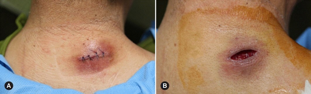
Fig. 3. Wound dehiscence due to postoperative surgical infection. (A) Before stitch out. (B) After stitch out.

Fig. 4. Histological findings of the epidermal cyst. (A) An arrow shows that the wall of the epidermal cyst is intact (hematoxylin and eosin stain, ×40). (B) An arrow shows that the ruptured wall of the epidermal cyst intact (hematoxylin and eosin stain, ×100).
Reference
-
References
1. Ogawa R. Benign and malignant nonmelanocytic tumors of the skin and soft tissue. In : Neligan PC, Warren RJ, Van Beek AL, editors. 3rd ed. London; New York: Elsevier Saunders;2013. p. 719–20.2. Min HJ, Lee JM, Han JK, Kim YJ. Influence factor in thickness of cyst wall of epidermal cysts. J Craniofac Surg. 2017; 28:e369–72.
Article3. Song SW, Burm JS, Yang WY, Kang SY. Minimally invasive excision of epidermal cysts through a small hole made by a CO2 laser. Arch Plast Surg. 2014; 41:85–8.
Article4. Yang HJ, Yang KC. A new method for facial epidermoid cyst removal with minimal incision. J Eur Acad Dermatol Venereol. 2009; 23:887–90.
Article5. Turk O, Ozdemir NG, Demirel N, Atci IB, Kanat A, Yolas C. Nontraumatic intradiploic epidermoid cyst and older age: association or causality? J Craniofac Surg. 2018; 29:e143–6.
Article
- Full Text Links
-
- Actions
-
Cited
- CITED
-
- Close
- Share
-
- Similar articles
-
- Molluscum contagiosum occuring in an epidermal cyst
- A Case of Hybrid Cyst: A Combined Epidermal and Trichilemmal Cyst
- A Case of Traumatic Epidermal Cyst Containing Glass Pieces
- A Case of a Follicular Hybrid Cyst (Epidermal Cyst and Pilomatricoma)
- Coexistence of Pilomatricoma and Epidermal Cyst (Hybrid Cyst)
