Yeungnam Univ J Med.
2019 May;36(2):85-91. 10.12701/yujm.2019.00136.
Endoscopic features aiding the diagnosis of gastric mucosa-associated lymphoid tissue lymphoma
- Affiliations
-
- 1Division of Gastroenterology and Hepatology, Department of Internal Medicine, Yeungnam University College of Medicine, Daegu, Korea. dr9696@gmail.com
- KMID: 2449319
- DOI: http://doi.org/10.12701/yujm.2019.00136
Abstract
- The incidence of gastric mucosa-associated lymphoid tissue (MALT) lymphoma is increasing worldwide, but the diagnosis is difficult. Most patients are asymptomatic or complain of nonspecific gastrointestinal symptoms. As the endoscopic features of gastric MALT lymphoma are variable and nonspecific, the possibility of this condition may be overlooked during esophagogastroduodenoscopy, and it remain undiagnosed. Therefore, this condition needs to be considered when an abnormal mucosa is observed during this procedure. Biopsy performed during endoscopy is the primary diagnostic test, but false negative results are possible; large numbers of samples should be collected from both normal and abnormal mucosae. Endoscopic ultrasonography is useful to assess the depth of invasion and to predict the treatment response. After treatment, follow-up tests are required every 3 months until complete remission is achieved, and annually thereafter. Early diagnosis of gastric MALT lymphoma is difficult, and its diagnosis and follow-up require wide experience and competent endoscopic technique.
Keyword
MeSH Terms
Figure
-
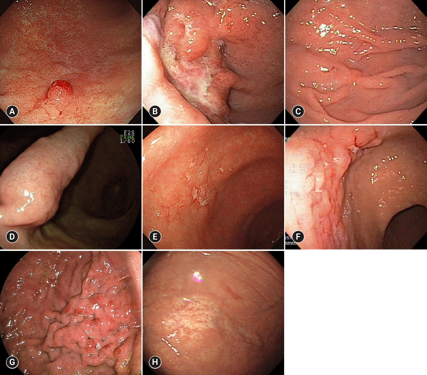
Fig. 1. Endoscopic findings of gastric mucosa-associated lymphoid tissue lymphoma. (A) Exophytic type. (B) Ulceroinfiltrative type. (C) IIc-like type. (D) Submucosal tumor type. (E) Multiple erosion type. (F) Cobblestone mucosa type. (G) Partial fold swelling type. (H) Discoloration type.
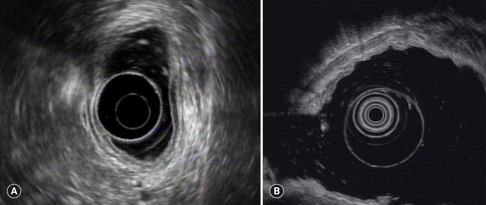
Fig. 2. Endoscopic ultrasonography findings of gastric mucosa-associated lymphoid tissue lymphoma. (A) Tumor limited to the mucosa. (B) Tumor invading the submucosa.

Fig. 3. Endoscopic findings of gastric MALT lymphoma after treatment. Mucosa, which was diagnosed as MALT lymphoma, had changed to atrophic and whitish discolored mucosa after Helicobacter pylori eradication. MALT, mucosa-associated lymphoid tissue.
Reference
-
References
1. Doglioni C, Wotherspoon AC, Moschini A, de Boni M, Isaacson PG. High incidence of primary gastric lymphoma in northeastern Italy. Lancet. 1992; 339:834–5.
Article2. Papaxoinis G, Papageorgiou S, Rontogianni D, Kaloutsi V, Fountzilas G, Pavlidis N, et al. Primary gastrointestinal non-Hodgkin's lymphoma: a clinicopathologic study of 128 cases in Greece. A Hellenic Cooperative Oncology Group study (HeCOG). Leuk Lymphoma. 2006; 47:2140–6.
Article3. Ferrucci PF, Zucca E. Primary gastric lymphoma pathogenesis and treatment: what has changed over the past 10 years? Br J Haematol. 2007; 136:521–38.4. Armitage JO. A clinical evaluation of the International Lymphoma Study Group classification of non-Hodgkin's lymphoma. The Non-Hodgkin's Lymphoma Classification Project. Blood. 1997; 89:3909–18.5. Olszewski AJ, Castillo JJ. Survival of patients with marginal zone lymphoma: analysis of the Surveillance, Epidemiology, and End Results database. Cancer. 2013; 119:629–38.6. Isaacson P, Wright DH. Malignant lymphoma of mucosa-associated lymphoid tissue. A distinctive type of B-cell lymphoma. Cancer. 1983; 52:1410–6.
Article7. Ruskoné-Fourmestraux A, Fischbach W, Aleman BM, Boot H, Du MQ, Megraud F, et al. EGILS consensus report. Gastric extranodal marginal zone B-cell lymphoma of MALT. Gut. 2011; 60:747–58.
Article8. Zucca E, Copie-Bergman C, Ricardi U, Thieblemont C, Raderer M, Ladetto Met al. Gastric marginal zone lymphoma of MALT type: ESMO Clinical Practice Guidelines for diagnosis, treatment and follow-up. Ann Oncol. 2013; 24(Suppl 6):vi144–8.
Article9. Zullo A, Hassan C, Cristofari F, Andriani A, De Francesco V, Ierardi E, et al. Effects of Helicobacter pylori eradication on early stage gastric mucosa-associated lymphoid tissue lymphoma. Clin Gastroenterol Hepatol. 2010; 8:105–10.
Article10. Ono S, Kato M, Takagi K, Kodaira J, Kubota K, Matsuno Y, et al. Long-term treatment of localized gastric marginal zone B-cell mucosa associated lymphoid tissue lymphoma including incidence of metachronous gastric cancer. J Gastroenterol Hepatol. 2010; 25:804–9.
Article11. Nakamura S, Sugiyama T, Matsumoto T, Iijima K, Ono S, Tajika M, et al. Long-term clinical outcome of gastric MALT lymphoma after eradication of Helicobacter pylori: a multicentre cohort follow-up study of 420 patients in Japan. Gut. 2012; 61:507–13.
Article12. Kelessis NG, Vassilopoulos PP, Tsamakidis KG, Bai MG, Avital S, Rosenthal RJ. Is gastroscopy still a valid diagnostic tool in detecting gastric MALT lymphomas? A dilemma beyond the eye. Mucosa-associated lymphoid tissue. Surg Endosc. 2003; 17:469–74.13. Stathis A, Chini C, Bertoni F, Proserpio I, Capella C, Mazzucchelli L, et al. Long-term outcome following Helicobacter pylori eradication in a retrospective study of 105 patients with localized gastric marginal zone B-cell lymphoma of MALT type. Ann Oncol. 2009; 20:1086–93.
Article14. Taal BG, den Hartog Jager FC, Tytgat GN. The endoscopic spectrum of primary non-Hodgkin's lymphoma of the stomach. Endoscopy. 1987; 19:190–2.
Article15. Zucca E, Bertoni F, Roggero E, Cavalli F. The gastric marginal zone B-cell lymphoma of MALT type. Blood. 2000; 96:410–9.
Article16. Lee SK, Lee YC, Chung JB, Chon CY, Moon YM, Kang JK, et al. Low grade gastric mucosa associated lymphoid tissue lymphoma: treatment strategies based on 10 year follow-up. World J Gastroenterol. 2004; 10:223–6.
Article17. Aoun JP, Moukarbel N, Khoury S. Endoscopic patterns of primary gastric MALT lymphoma. J Med Liban. 1998; 46:131–5.18. Yokoi T, Nakamura T, Nakamura S. Differential diagnosis of gastric MALT lymphomas. Stomach Intest. 2001; 36:13–20.19. Caletti G, Barbara L. Gastric lymphoma: difficult to diagnose, difficult to stage? Endoscopy. 1993; 25:528–30.
Article20. Taal BG, Boot H, van Heerde P, de Jong D, Hart AA, Burgers JM. Primary non-Hodgkin lymphoma of the stomach: endoscopic pattern and prognosis in low versus high grade malignancy in relation to the MALT concept. Gut. 1996; 39:556–61.
Article21. Hu Q, Zhang Y, Zhang X, Fu K. Gastric mucosa-associated lymphoid tissue lymphoma and Helicobacter pylori infection: a review of current diagnosis and management. Biomark Res. 2016; 4:15.
Article22. Suekane H, Iida M, Kuwano Y, Kohrogi N, Yao T, Iwashita A, et al. Diagnosis of primary early gastric lymphoma. Usefulness of endoscopic mucosal resection for histologic evaluation. Cancer. 1993; 71:1207–13.
Article23. Wotherspoon AC, Doglioni C, Diss TC, Pan L, Moschini A, de Boni M, et al. Regression of primary low-grade B-cell gastric lymphoma of mucosa-associated lymphoid tissue type after eradication of Helicobacter pylori. Lancet. 1993; 342:575–7.
Article24. Hummel M, Oeschger S, Barth TF, Loddenkemper C, Cogliatti SB, Marx A, et al. Wotherspoon criteria combined with B cell clonality analysis by advanced polymerase chain reaction technology discriminates covert gastric marginal zone lymphoma from chronic gastritis. Gut. 2006; 55:782–7.
Article25. Du MQ, Atherton JC. Molecular subtyping of gastric MALT lymphomas: implications for prognosis and management. Gut. 2006; 55:886–93.
Article26. Liu H, Ruskon-Fourmestraux A, Lavergne-Slove A, Ye H, Molina T, Bouhnik Y, et al. Resistance of t(11;18) positive gastric mucosa-associated lymphoid tissue lymphoma to Helicobacter pylori eradication therapy. Lancet. 2001; 357:39–40.
Article27. Zullo A, Hassan C, Andriani A, Cristofari F, Cardinale V, Spinelli GP, et al. Primary low-grade and high-grade gastric MALT-lymphoma presentation. J Clin Gastroenterol. 2010; 44:340–4.
Article28. Malfertheiner P, Megraud F, O'Morain CA, Gisbert JP, Kuipers EJ, Axon AT, et al. Management of Helicobacter pylori infection-the Maastricht V/Florence Consensus Report. Gut. 2017; 66:6–30.
Article29. Hartman DJ, Owens SR. Are routine ancillary stains required to diagnose Helicobacter infection in gastric biopsy specimens? An institutional quality assurance review. Am J Clin Pathol. 2012; 137:255–60.
Article30. Wang XI, Zhang S, Abreo F, Thomas J. The role of routine immunohistochemistry for Helicobacter pylori in gastric biopsy. Ann Diagn Pathol. 2010; 14:256–9.
Article31. Nonaka K, Ishikawa K, Arai S, Nakao M, Shimizu M, Sakurai T, et al. A case of gastric mucosa-associated lymphoid tissue lymphoma in which magnified endoscopy with narrow band imaging was useful in the diagnosis. World J Gastrointest Endosc. 2012; 4:151–6.
Article32. Deng P, Min M, Ma CY, Liu Y. Linked color imaging technology for diagnosis of gastric mucosa-associated lymphoid tissue lymphoma. Chin Med J (Engl). 2017; 130:2759–60.
Article33. Caletti G, Ferrari A, Brocchi E, Barbara L. Accuracy of endoscopic ultrasonography in the diagnosis and staging of gastric cancer and lymphoma. Surgery. 1993; 113:14–27.34. Palazzo L, Roseau G, Ruskone-Fourmestraux A, Rougier P, Chaussade S, Rambaud JC, et al. Endoscopic ultrasonography in the local staging of primary gastric lymphoma. Endoscopy. 1993; 25:502–8.
Article35. Mehra M, Agarwal B. Endoscopic diagnosis and staging of mucosa-associated lymphoid tissue lymphoma. Curr Opin Gastroenterol. 2008; 24:623–6.
Article36. Lévy M, Hammel P, Lamarque D, Marty O, Chaumette MT, Haioun C, et al. Endoscopic ultrasonography for the initial staging and follow-up in patients with low-grade gastric lymphoma of mucosa-associated lymphoid tissue treated medically. Gastrointest Endosc. 1997; 46:328–33.
Article37. Queneau PE, Helg C, Brundler MA, Frossard JL, Spahr L, Girardet C, et al. Diagnosis of a gastric mucosa-associated lymphoid tissue lymphoma by endoscopic ultrasonography-guided biopsies in a patient with a parotid gland localization. Scand J Gastroenterol. 2002; 37:493–6.
Article38. El-Zahabi LM, Jamali FR, El-Hajj II, Naja M, Salem Z, Shamseddine A, et al. The value of EUS in predicting the response of gastric mucosa-associated lymphoid tissue lymphoma to Helicobacter pylori eradication. Gastrointest Endosc. 2007; 65:89–96.
Article39. Sheu BS, Shiesh SC, Wang JT, Yang HB, Lin ST, Wu JJ. Clinical application of 20 MHz endosonography and anti-Helicobacter pylori immunoblots to predict regression of low-grade gastric MALToma by H. pylori eradication. Helicobacter. 2003; 8:36–45.
Article40. Yeh HZ, Chen GH, Chang WD, Poon SK, Yang SS, Lien HC, et al. Long-term follow up of gastric low-grade mucosa-associated lymphoid tissue lymphoma by endosonography emphasizing the application of a miniature ultrasound probe. J Gastroenterol Hepatol. 2003; 18:162–7.
Article41. Janssen J. The impact of EUS in primary gastric lymphoma. Best Pract Res Clin Gastroenterol. 2009; 23:671–8.
Article42. Choi YJ, Lee DH, Kim JY, Kwon JE, Kim JY, Jo HJ, et al. Low grade gastric mucosa-associated lymphoid tissue lymphoma: clinicopathological factors associated with helicobacter pylori eradication and tumor regression. Clin Endosc. 2011; 44:101–8.
Article43. Ryu KD, Kim GH, Park SO, Lee KJ, Moon JY, Jeon HK, et al. Treatment outcome for gastric mucosa-associated lymphoid tissue lymphoma according to Helicobacter pylori infection status: a single-center experience. Gut Liver. 2014; 8:408–14.
Article44. Thieblemont C, Zucca E. Clinical aspects and therapy of gastrointestinal MALT lymphoma. Best Pract Res Clin Haematol. 2017; 30:109–17.
Article45. Wündisch T, Thiede C, Morgner A, Dempfle A, Günther A, Liu H, et al. Long-term follow-up of gastric MALT lymphoma after Helicobacter pylori eradication. J Clin Oncol. 2005; 23:8018–24.46. Urakami Y, Sano T, Begum S, Endo H, Kawamata H, Oki Y. Endoscopic characteristics of low-grade gastric mucosa-associated lymphoid tissue lymphoma after eradication of Helicobacter pylori. J Gastroenterol Hepatol. 2000; 15:1113–9.
Article47. Ono S, Kato M, Ono Y, Nakagawa M, Shimizu Y, Asaka M. Magnified endoscopic images of gastric MALT lymphoma before and after treatment. Endoscopy. 2007; 39(Suppl 1):E328.
Article48. Ishihara R, Tatsuta M, Iishi H, Uedo N, Narahara H, Ishiguro S. Usefulness of endoscopic appearance for choosing a biopsy target site and determining complete remission of primary gastric lymphoma of mucosa-associated lymphoid tissue after eradication of Helicobacter pylori infection. Am J Gastroenterol. 2002; 97:772–4.
Article49. Palmela C, Fonseca C, Faria R, Baptista RB, Ribeiro S, Ferreira AO. Increased risk for metachronous gastric adenocarcinoma following gastric MALT lymphoma-A US population-based study. United European Gastroenterol J. 2017; 5:473–8.
Article
- Full Text Links
-
- Actions
-
Cited
- CITED
-
- Close
- Share
-
- Similar articles
-
- Endoscopic Diagnosis of Gastric MALT Lymphoma
- A Polypoid Mucosa-Associated Lymphoid Tissue Lymphoma of the Stomach Treated with Endoscopic Polypectomy
- Two Cases of Low-Grade Gastric Mucosa Associated Lymphoid Tissue Lymphoma with Normal Endoscopic Appearance
- Successful Endoscopic Resection of Gastric Mucosa-Associated Lymphoid Tissue Lymphoma Unresponsive to Helicobacter pylori Eradication Therapy
- Role of Chemotherapy in Gastric Marginal Zone B-Cell Lymphoma of Mucosa-Associated Lymphoid Tissue (MALT) Type
