Evaluation of Validity and Reliability of Inertial Measurement Unit-Based Gait Analysis Systems
- Affiliations
-
- 1Department of Rehabilitation Medicine, Hanyang University College of Medicine, Seoul, Korea. msb1220@hanmail.net
- 2Department of Arts & Technology, School of Industrial Information Studies, Hanyang University, Seoul, Korea.
- KMID: 2448909
- DOI: http://doi.org/10.5535/arm.2018.42.6.872
Abstract
OBJECTIVE
To replace camera-based three-dimensional motion analyzers which are widely used to analyze body movements and gait but are also costly and require a large dedicated space, this study evaluates the validity and reliability of inertial measurement unit (IMU)-based systems by analyzing their spatio-temporal and kinematic measurement parameters.
METHODS
The investigation was conducted in three separate hospitals with three healthy participants. IMUs were attached to the abdomen as well as the thigh, shank, and foot of both legs of each participant. Each participant then completed a 10-m gait course 10 times. During each gait cycle, the hips, knees, and ankle joints were observed from the sagittal, frontal, and transverse planes. The experiments were conducted with both a camerabased system and an IMU-based system. The measured gait analysis data were evaluated for validity and reliability using root mean square error (RMSE) and intraclass correlation coefficient (ICC) analyses.
RESULTS
The differences between the RMSE values of the two systems determined through kinematic parameters ranged from a minimum of 1.83 to a maximum of 3.98 with a tolerance close to 1%. The results of this study also confirmed the reliability of the IMU-based system, and all of the variables showed a statistically high ICC.
CONCLUSION
These results confirmed that IMU-based systems can reliably replace camera-based systems for clinical body motion and gait analyses.
MeSH Terms
Figure
-
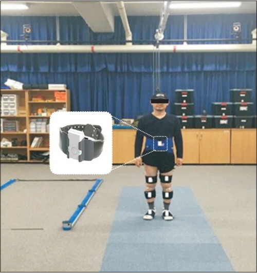
Fig. 1. A subject has inertial measurement units on both dorsa of the feet, shafts of the tibias, middles of the femurs, and the lower abdomen in the room where the camera-based system is installed.
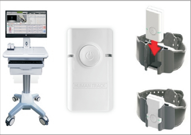
Fig. 2. The inertial measurement unit sensor is placed on a holder so as to increase stability and accuracy.
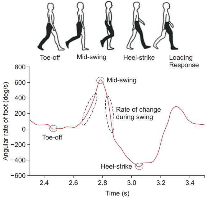
Fig. 3. The angle of rotation of the foot was used to determine the gait event through a gyroscope on the foot.
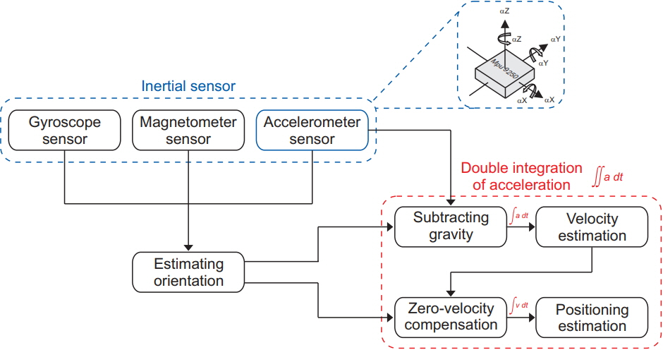
Fig. 4. A two-phase cumulative error reduction algorithm is used to minimize the accumulated error of the double integration, which was calculated using the acceleration values and the angular velocity from the attitude and heading reference system (AHRS) modules.
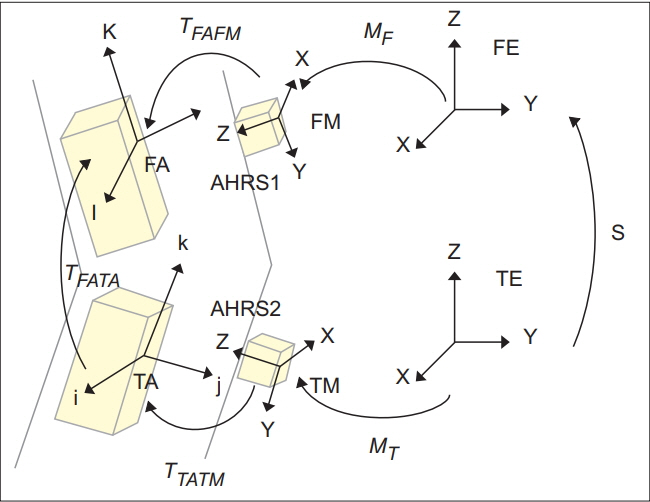
Fig. 5. A conceptual map presents the segmental joint angle calculation method in the knee joint as an example. The same algorithm can be applied to other segmental joint angles. AHRS, attitude and heading reference system; TA, tibia anatomical; TM, tibia measurement; FA, femur anatomical; FM, femur measurement.
Cited by 2 articles
-
Novel Method of Classification in Knee Osteoarthritis: Machine Learning Application Versus Logistic Regression Model
Jung Ho Yang, Jae Hyeon Park, Seong-Ho Jang, Jaesung Cho
Ann Rehabil Med. 2020;44(6):415-427. doi: 10.5535/arm.20071.Determining the Most Appropriate Assistive Walking Device Using the Inertial Measurement Unit-Based Gait Analysis System in Disabled Patients
Junhee Lee, Chang Hoon Bae, Aeri Jang, Seoyon Yang, Hasuk Bae
Ann Rehabil Med. 2020;44(1):48-57. doi: 10.5535/arm.2020.44.1.48.
Reference
-
1. Simon SR. Quantification of human motion: gait analysis-benefits and limitations to its application to clinical problems. J Biomech. 2004; 37:1869–80.
Article2. Sutherland DH. The evolution of clinical gait analysis. Part II. kinematics. Gait Posture. 2002; 16:159–79.3. Sutherland DH. The evolution of clinical gait analysis. Part III. Kinetics and energy assessment. Gait Posture. 2005; 21:447–61.4. Ugbolue UC, Papi E, Kaliarntas KT, Kerr A, Earl L, Pomeroy VM, et al. The evaluation of an inexpensive, 2D, video based gait assessment system for clinical use. Gait Posture. 2013; 38:483–9.
Article5. Costigan PA, Deluzio KJ, Wyss UP. Knee and hip kinetics during normal stair climbing. Gait Posture. 2002; 16:31–7.
Article6. Favre J, Jolles BM, Aissaoui R, Aminian K. Ambulatory measurement of 3D knee joint angle. J Biomech. 2008; 41:1029–35.
Article7. Watanabe T, Saito H, Koike E, Nitta K. A preliminary test of measurement of joint angles and stride length with wireless inertial sensors for wearable gait evaluation system. Comput Intell Neurosci. 2011; 2011:975193.
Article8. Zheng H, Black ND, Harris ND. Position-sensing technologies for movement analysis in stroke rehabilitation. Med Biol Eng Comput. 2005; 43:413–20.
Article9. Cloete T, Scheffer C. Benchmarking of a full-body inertial motion capture system for clinical gait analysis. Conf Proc IEEE Eng Med Biol Soc. 2008; 2008:4579–82.
Article10. Benedetti MG, Merlo A, Leardini A. Inter-laboratory consistency of gait analysis measurements. Gait Posture. 2013; 38:934–9.
Article11. Boudarham J, Roche N, Pradon D, Bonnyaud C, Bensmail D, Zory R. Variations in kinematics during clinical gait analysis in stroke patients. PLoS One. 2013; 8:e66421.
Article12. Karasawa Y, Teruyama Y, Watanabe T. A trial of making reference gait data for simple gait evaluation system with wireless inertial sensors. Conf Proc IEEE Eng Med Biol Soc. 2013; 2013:3427–30.
Article13. van Acht V, Bongers E, Lambert N, Verberne R. Miniature wireless inertial sensor for measuring human motions. Conf Proc IEEE Eng Med Biol Soc. 2007; 2007:6279–82.
Article14. Luinge HJ, Veltink PH. Measuring orientation of human body segments using miniature gyroscopes and accelerometers. Med Biol Eng Comput. 2005; 43:273–82.
Article15. Zhu R, Zhou Z. A real-time articulated human motion tracking using tri-axis inertial/magnetic sensors package. IEEE Trans Neural Syst Rehabil Eng. 2004; 12:295–302.16. Kim MS, Yu SB, Lee KS. Development of a high-precision calibration method for inertial measurement unit. Int J Precis Eng Manuf. 2014; 15:567–75.
Article17. Lee D, Lee S, Park S, Ko S. Test and error parameter estimation for MEMS—based low cost IMU calibration. Int J Precis Eng Manuf. 2011; 12:597–603.18. Madgwick S. An efficient orientation filter for inertial and inertial/magnetic sensor arrays [Internet]. Bristol, UK: X-IO Technologies;2010. [cited 2018 Nov 15]. Available from: http://x-io.co.uk/res/doc/madgwick_internal_report.pdf.19. Alvarez JC, Gonzalez RC, Alvarez D, Lopez AM, Rodriguez-Uria J. Multisensor approach to walking distance estimation with foot inertial sensing. Conf Proc IEEE Eng Med Biol Soc. 2007; 2007:5720–3.
Article20. Dejnabadi H, Jolles BM, Aminian K. A new approach to accurate measurement of uniaxial joint angles based on a combination of accelerometers and gyroscopes. IEEE Trans Biomed Eng. 2005; 52:1478–84.
Article21. Lau H, Tong K. The reliability of using accelerometer and gyroscope for gait event identification on persons with dropped foot. Gait Posture. 2008; 27:248–57.
Article22. Tong K, Granat MH. A practical gait analysis system using gyroscopes. Med Eng Phys. 1999; 21:87–94.
Article23. Brennan A, Zhang J, Deluzio K, Li Q. Quantification of inertial sensor-based 3D joint angle measurement accuracy using an instrumented gimbal. Gait Posture. 2011; 34:320–3.
Article24. Cho JS, Kang SI, Lee KH, Jang SH, Kim IY, Lee JS. Implementation of gait analysis system based on inertial sensors. J Rehabil Welf Eng Assist Technol. 2015; 9:137–44.25. Patterson MR, Delahunt E, Sweeney KT, Caulfield B. An ambulatory method of identifying anterior cruciate ligament reconstructed gait patterns. Sensors (Basel). 2014; 14:887–99.
Article26. Greene BR, McGrath D, O’Neill R, O’Donovan KJ, Burns A, Caulfield B. An adaptive gyroscope-based algorithm for temporal gait analysis. Med Biol Eng Comput. 2010; 48:1251–60.
Article27. Salarian A, Russmann H, Vingerhoets FJ, Dehollain C, Blanc Y, Burkhard PR, et al. Gait assessment in Parkinson’s disease: toward an ambulatory system for longterm monitoring. IEEE Trans Biomed Eng. 2004; 51:1434–43.
Article28. Sabatini AM, Martelloni C, Scapellato S, Cavallo F. Assessment of walking features from foot inertial sensing. IEEE Trans Biomed Eng. 2005; 52:486–94.
Article29. Feliz Alonso R, Zalama Casanova E, Gomez Garcia-Bermejo J. Pedestrian tracking using inertial sensors. J Phys Agents. 2009; 3:35–43.
Article30. Lee KH, Kang SI, Cho JS, Lim DH, Lee JS, Kim IY. Development of gait distance measurement system based on inertial measurement units. J Rehabil Welf Eng Assist Technol. 2015; 9:161–8.31. Zhou H, Hu H. Inertial sensors for motion detection of human upper limbs. Sens Rev. 2007; 27:151–8.
Article32. Kim CY, Hong JS, Chun KJ. Validation of feasibility of two depth sensor-based Microsoft Kinect cameras for human abduction-adduction motion analysis. Int J Precis Eng Manuf. 2016; 17:1209–14.
Article33. Hamacher D, Hamacher D, Taylor WR, Singh NB, Schega L. Towards clinical application: repetitive sensor position re-calibration for improved reliability of gait parameters. Gait Posture. 2014; 39:1146–8.
Article34. Lutzner C, Voigt H, Roeder I, Kirschner S, Lutzner J. Placement makes a difference: accuracy of an accelerometer in measuring step number and stair climbing. Gait Posture. 2014; 39:1126–32.35. Welch G, Foxlin E. Motion tracking: no silver bullet, but a respectable arsenal. IEEE Comput Graph Appl. 2002; 22:24–38.
Article36. Bautmans I, Jansen B, Van Keymolen B, Mets T. Reliability and clinical correlates of 3D-accelerometry based gait analysis outcomes according to age and fall-risk. Gait Posture. 2011; 33:366–72.
Article37. Henriksen M, Lund H, Moe-Nilssen R, Bliddal H, Danneskiod-Samsoe B. Test-retest reliability of trunk accelerometric gait analysis. Gait Posture. 2004; 19:288–97.
Article38. Prince B, Makrides L, Richman J. Research methodology and applied statistics. Part 2. The literature search. Physiother Can. 1980; 32:201–6.
- Full Text Links
-
- Actions
-
Cited
- CITED
-
- Close
- Share
-
- Similar articles
-
- Determining the Most Appropriate Assistive Walking Device Using the Inertial Measurement Unit-Based Gait Analysis System in Disabled Patients
- Validity and Reliability of an Inertial Measurement Unit-Based 3D Angular Measurement of Shoulder Joint Motion
- Validation of foot pitch angle estimation using inertial measurement unit against marker-based optical 3D motion capture system
- Comparison of Gait Parameters during Forward Walking under Different Visual Conditions Using Inertial Motion Sensors
- Quantitative Analysis of Gait and Balance
