Treatment of Exposed Hydroxyapatite Orbital Implants Wrapped with a Synthetic Dura Substitute
- Affiliations
-
- 1Department of Ophthalmology, Institute of Vision Research, Severance Hospital, Yonsei University College of Medicine, Seoul, Korea. yoonjs@yuhs.ac
- KMID: 2448866
- DOI: http://doi.org/10.3341/kjo.2019.0018
Abstract
- PURPOSE
To describe cases of exposed hydroxyapatite (HA) implants wrapped with the synthetic dura substitute Neuro-Patch treated via simple Neuro-Patch removal.
METHODS
The medical records of seven patients who experienced exposure of their HA implant were reviewed. All patients had been enucleated and implanted with HA wrapped with Neuro-Patch. For treatment, Neuro-Patch was removed to the greatest extent possible. After applying local anesthesia with lidocaine, blunt dissection was performed to separate the conjunctiva and Neuro-Patch via the site of exposure. Pressure was applied to the remaining Neuro-Patch with forceps and removed with scissors.
RESULTS
Neuro-Patch was visible at the area of exposure in all patients. No surgery beyond initial Neuro-Patch removal was necessary in six of the seven patients. In five cases, the exposed area began to heal rapidly after Neuro-patch removal without primary closure of the defect. In one case, the Neuro-Patch material and all necrotic tissue was removed aggressively due to inflammation around the orbital implant. Lastly, an infection was noted in one case, prompting complete removal of the Neuro-Patch-wrapped HA implant.
CONCLUSIONS
Wrapping material may hinder implant vascularization. Exposure of HA in wrapped implants can be successfully treated by a simple removal procedure if detected and managed early.
MeSH Terms
Figure
-

Fig. 1 Anophthalmic sockets in cases 1 to 6. (A) Case 1, (B) case 2, (C) case 3, (D) case 4, (E) case 5, and (F) case 6. The upper images in panels show areas of exposed Neuro-Patch of various sizes and lower images show well healed areas of former exposure after Neuro-Patch removal. The formerly exposed areas remained well healed at the last follow-up examination in all six cases. Written informed consent from the patient was obtained.

Fig. 2 Case 6. (A) Preoperative computed tomography showing inflammation around the orbital implant (arrow). (B) Postoperative computed tomography showing remarkable improvement of the inflammation around the orbital implant. Written informed consent from the patient was obtained.
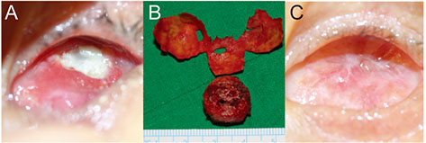
Fig. 3 Case 7. Infected Neuro-Patch–wrapped implant. (A) Neuro-Patch exposure with severe discharge due to implant infection, (B) removed hydroxyapatite implant and Neuro-Patch, (C) well healed conjunctival socket postoperatively. Written informed consent from the patient was obtained.
Reference
-
1. Dutton JJ. Coralline hydroxyapatite as an ocular implant. Ophthalmology. 1991; 98:370–377.
Article2. Perry AC. Integrated orbital implants. Adv Ophthalmic Plast Reconstr Surg. 1990; 8:75–81.
Article3. Jordan DR, Klapper SR. Wrapping hydroxyapatite implants. Ophthalmic Surg Lasers. 1999; 30:403–407.4. Lee V, Subak-Sharpe I, Hungerford JL, et al. Exposure of primary orbital implants in postenucleation retinoblastoma patients. Ophthalmology. 2000; 107:940–945.5. Shields CL, Shields JA, De Potter P. Hydroxyapatite orbital implant after enucleation. Experience with initial 100 consecutive cases. Arch Ophthalmol. 1992; 110:333–338.
Article6. Shields JA, Shields CL, De Potter P. Enucleation technique for children with retinoblastoma. J Pediatr Ophthalmol Strabismus. 1992; 29:213–215.
Article7. Yamada S, Aiba T, Endo Y, et al. Creutzfeldt-Jakob disease transmitted by a cadaveric dura mater graft. Neurosurgery. 1994; 34:740–743.
Article8. Mehta JS, Franks WA. The sclera, the prion, and the ophthalmologist. Br J Ophthalmol. 2002; 86:587–592.
Article9. Raul JS, Godard J, Arbez-Gindre F, Czorny A. Use of polyester urethane (Neuro-Patch) as a dural substitute. Prospective study of 70 cases. Neurochirurgie. 2003; 49(2-3 Pt 1):83–89.10. Seiff SR, Chang JS Jr, Hurt MH, Khayam-Bashi H. Polymerase chain reaction identification of human immunodeficiency virus-1 in preserved human sclera. Am J Ophthalmol. 1994; 118:528–530.
Article11. Simonds RJ, Holmberg SD, Hurwitz RL, et al. Transmission of human immunodeficiency virus type 1 from a seronegative organ and tissue donor. N Engl J Med. 1992; 326:726–732.
Article12. Jordan DR, Ells A, Brownstein S, et al. Vicryl-mesh wrap for the implantation of hydroxyapatite orbital implants: an animal model. Can J Ophthalmol. 1995; 30:241–246.13. Jordan DR, Allen LH, Ells A, et al. The use of Vicryl mesh (polyglactin 910) for implantation of hydroxyapatite orbital implants. Ophthalmic Plast Reconstr Surg. 1995; 11:95–99.
Article14. Oestreicher JH, Liu E, Berkowitz M. Complications of hydroxyapatite orbital implants. A review of 100 consecutive cases and a comparison of Dexon mesh (polyglycolic acid) with scleral wrapping. Ophthalmology. 1997; 104:324–329.15. Gudmundsson G, Sogaard I. Complications to the use of vicryl-collagen dural substitute. Acta Neurochir (Wien). 1995; 132:145–147.
Article16. Malliti M, Page P, Gury C, et al. Comparison of deep wound infection rates using a synthetic dural substitute (neuro-patch) or pericranium graft for dural closure: a clinical review of 1 year. Neurosurgery. 2004; 54:599–603.
Article17. El Majdoub F, Lohr M, Maarouf M, et al. Transmigration of fibrino-purulent inflammation and malignant cells into an artificial dura substitute (Neuro-Patch): report of two cases. Acta Neurochir (Wien). 2009; 151:833–835.
Article18. Heimann H, Bechrakis NE, Zepeda LC, et al. Exposure of orbital implants wrapped with polyester-urethane after enucleation for advanced retinoblastoma. Ophthalmic Plast Reconstr Surg. 2005; 21:123–128.
Article19. Kaltreider SA, Newman SA. Prevention and management of complications associated with the hydroxyapatite implant. Ophthalmic Plast Reconstr Surg. 1996; 12:18–31.
Article
- Full Text Links
-
- Actions
-
Cited
- CITED
-
- Close
- Share
-
- Similar articles
-
- Histopathologic Comparison of Vascularization between Dacron and Donor Sclera as Wrapping Material in Hydroxyapatite Implantation
- Management of Exposed Porous Orbital Implant with Autogenous Dermis Graft
- Management of Exposed Hydroxyapatite Implant with Acellular Dermal Allograft
- Tube Erosion with Scleral Melting after Ahmed Valve Implantation Using a Synthetic Dural Substitute
- Hydroxyapatite Implantation using Autogenous Temporalis Muscle Fascia
