Analysis of Circular RNAs in the Coronary Arteries of Patients with Kawasaki Disease
- Affiliations
-
- 1Department of Biochemistry, Chonnam National University Medical School, Hwasun, Korea. ykk@jnu.ac.kr
- KMID: 2447756
- DOI: http://doi.org/10.12997/jla.2019.8.1.50
Keyword
Figure
-
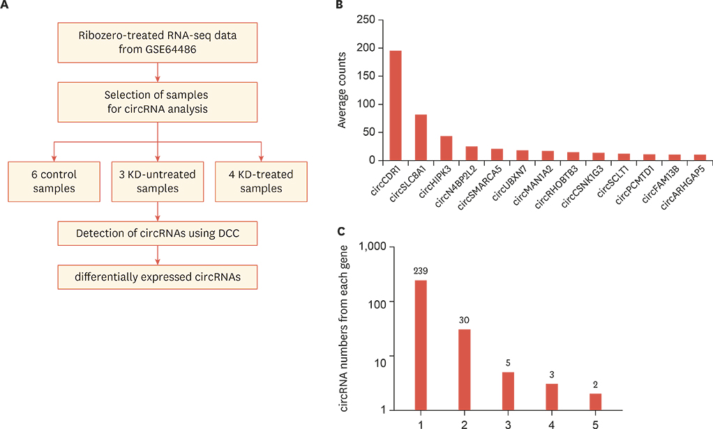
Fig. 1 Identification of circRNAs expressed in coronary artery tissue from control and Kawasaki disease groups. (A) The procedure used to identify circRNAs in the control and Kawasaki disease samples. Based on data from the GSE64486 dataset, the expression level of circRNAs in each group was calculated using the DCC algorithm and differentially expressed circRNAs were selected for further study.1316 (B) Average expression counts of highly expressed circRNAs. circRNAs with normalized average counts greater than 10 are shown. (C) The number of circRNAs produced from each gene was calculated. circRNA, circular RNA.
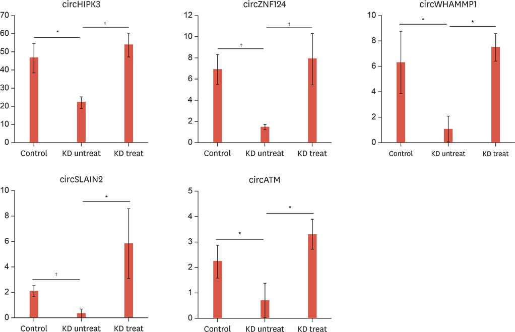
Fig. 2 Selected circRNAs with probable roles in Kawasaki disease. Among the circRNAs listed in Supplementary Table 2, only circRNAs that showed differential expression between control and untreated Kawasaki disease groups (p<0.05), and between untreated and treated Kawasaki disease groups (p<0.05), but no significant difference between the control and treated Kawasaki disease groups (p>0.05), were selected. p values were calculated by a t-test between samples from 2 groups, respectively. circRNA, circular RNA. *p<0.05; †p<0.01.
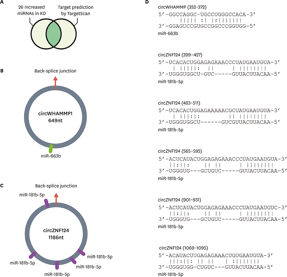
Fig. 3 Possible working mechanism of circRNAs as miRNA suppressors. (A) Analysis of circRNAs as miRNA suppressors. Among the increased miRNAs in patients with Kawasaki disease, miRNAs with predicted binding sites in circRNA sequences were selected using TargetScan.19 (B-C) The miRNA binding sites in circRNAs are depicted for (B) circWHAMMP1 and (C) circZNF124. (D) Base pairing between the predicted miRNA binding sites and circRNAs is shown. The nucleotide position of the back-splice junction depicted in (B, C) was set as zero, and the relative position of the nucleotide sequences was calculated. circRNA, circular RNA; miRNA, microRNA.
Cited by 1 articles
-
Circular RNA as a Possible Novel Biomarker for Kawasaki Disease
Kyung Lim Yoon
J Lipid Atheroscler. 2019;8(1):48-49. doi: 10.12997/jla.2019.8.1.48.
Reference
-
1. Newburger JW, Takahashi M, Burns JC. Kawasaki disease. J Am Coll Cardiol. 2016; 67:1738–1749.
Article2. Cohen E, Sundel R. Kawasaki disease at 50 years. JAMA Pediatr. 2016; 170:1093–1099.
Article3. Uehara R, Belay ED. Epidemiology of Kawasaki disease in Asia, Europe, and the United States. J Epidemiol. 2012; 22:79–85.
Article4. Ha S, Seo GH, Kim KY, Kim DS. Epidemiologic study on Kawasaki disease in Korea, 2007–2014: based on Health Insurance Review & Assessment Service Claims. J Korean Med Sci. 2016; 31:1445–1449.
Article5. Newburger JW, Takahashi M, Gerber MA, Gewitz MH, Tani LY, Burns JC, et al. Diagnosis, treatment, and long-term management of Kawasaki disease: a statement for health professionals from the Committee on Rheumatic Fever, Endocarditis and Kawasaki Disease, Council on Cardiovascular Disease in the Young, American Heart Association. Circulation. 2004; 110:2747–2771.
Article6. Glažar P, Papavasileiou P, Rajewsky N. circBase: a database for circular RNAs. RNA. 2014; 20:1666–1670.
Article7. Ebbesen KK, Kjems J, Hansen TB. Circular RNAs: identification, biogenesis and function. Biochim Biophys Acta. 2016; 1859:163–168.
Article8. Wilusz JE. Circular RNAs: unexpected outputs of many protein-coding genes. RNA Biol. 2017; 14:1007–1017.
Article9. Kulcheski FR, Christoff AP, Margis R. Circular RNAs are miRNA sponges and can be used as a new class of biomarker. J Biotechnol. 2016; 238:42–51.
Article10. Du WW, Zhang C, Yang W, Yong T, Awan FM, Yang BB. Identifying and characterizing circRNA-protein interaction. Theranostics. 2017; 7:4183–4191.
Article11. Pamudurti NR, Bartok O, Jens M, Ashwal-Fluss R, Stottmeister C, Ruhe L, et al. Translation of CircRNAs. Mol Cell. 2017; 66:9–21.e7.
Article12. Legnini I, Di Timoteo G, Rossi F, Morlando M, Briganti F, Sthandier O, et al. Circ-ZNF609 is a circular RNA that can be translated and functions in myogenesis. Mol Cell. 2017; 66:22–37.e9.
Article13. Rowley AH, Wylie KM, Kim KY, Pink AJ, Yang A, Reindel R, et al. The transcriptional profile of coronary arteritis in Kawasaki disease. BMC Genomics. 2015; 16:1076.
Article14. Bolger AM, Lohse M, Usadel B. Trimmomatic: a flexible trimmer for Illumina sequence data. Bioinformatics. 2014; 30:2114–2120.
Article15. Dobin A, Davis CA, Schlesinger F, Drenkow J, Zaleski C, Jha S, et al. STAR: ultrafast universal RNA-seq aligner. Bioinformatics. 2013; 29:15–21.
Article16. Cheng J, Metge F, Dieterich C. Specific identification and quantification of circular RNAs from sequencing data. Bioinformatics. 2016; 32:1094–1096.
Article17. Tyner C, Barber GP, Casper J, Clawson H, Diekhans M, Eisenhart C, et al. The UCSC Genome Browser database: 2017 update. Nucleic Acids Res. 2017; 45:D626–D634.18. Rowley AH, Pink AJ, Reindel R, Innocentini N, Baker SC, Shulman ST, et al. A study of cardiovascular miRNA biomarkers for Kawasaki disease. Pediatr Infect Dis J. 2014; 33:1296–1299.
Article19. Agarwal V, Bell GW, Nam JW, Bartel DP. Predicting effective microRNA target sites in mammalian mRNAs. eLife. 2015; 4:e05005.
Article20. Memczak S, Jens M, Elefsinioti A, Torti F, Krueger J, Rybak A, et al. Circular RNAs are a large class of animal RNAs with regulatory potency. Nature. 2013; 495:333–338.
Article21. Hansen TB, Jensen TI, Clausen BH, Bramsen JB, Finsen B, Damgaard CK, et al. Natural RNA circles function as efficient microRNA sponges. Nature. 2013; 495:384–388.
Article22. Clough E, Barrett T. The gene expression omnibus database. Methods Mol Biol. 2016; 1418:93–110.
Article23. Shan K, Liu C, Liu BH, Chen X, Dong R, Liu X, et al. Circular noncoding RNA HIPK3 mediates retinal vascular dysfunction in diabetes mellitus. Circulation. 2017; 136:1629–1642.
Article24. Zheng Q, Bao C, Guo W, Li S, Chen J, Chen B, et al. Circular RNA profiling reveals an abundant circHIPK3 that regulates cell growth by sponging multiple miRNAs. Nat Commun. 2016; 7:11215.
Article25. Liang S, Zhang N, Deng Y, Chen L, Zhang Y, Zheng Z, et al. miR-663b promotes tumor cell proliferation, migration and invasion in nasopharyngeal carcinoma through targeting TUSC2. Exp Ther Med. 2017; 14:1095–1103.
Article26. Hori D, Dunkerly-Eyring B, Nomura Y, Biswas D, Steppan J, Henao-Mejia J, et al. miR-181b regulates vascular stiffness age dependently in part by regulating TGF-β signaling. PLoS One. 2017; 12:e0174108.
Article
- Full Text Links
-
- Actions
-
Cited
- CITED
-
- Close
- Share
-
- Similar articles
-
- Kawasaki Disease
- CABG for an Adult with Coronary Disease due to Kawasaki Disease
- Changes in Coronary Perfusion after Occlusion of Coronary Arteries in Kawasaki Disease
- Three cases of atypical Kawasaki disease with coronary aneurysm
- A Clinical Observation of Kawasaki Disease at High Risk of Coronary Artery Aneurysm
