Clin Exp Otorhinolaryngol.
2019 May;12(2):224-230. 10.21053/ceo.2018.01011.
Role of Immunohistochemistry in Fine Needle Aspiration and Core Needle Biopsy of Thyroid Nodules
- Affiliations
-
- 1Department of Otorhinolaryngology-Head and Neck Surgery, Seoul National University Hospital, Seoul, Korea. ahnsh30@snu.ac.kr
- 2Department of Pathology, Seoul National University Bundang Hospital, Seoul National University College of Medicine, Seongnam, Korea.
- KMID: 2447433
- DOI: http://doi.org/10.21053/ceo.2018.01011
Abstract
OBJECTIVES
Immunohistochemistry (IHC) has been used for the diagnosis of indeterminate results in fine needle aspiration (FNA) of thyroid nodules. However, the role of IHC in core needle biopsy (CNB) is not clear and the efficacy of testing for molecular markers following CNB has not been evaluated. The aim of this study is to compare the role of IHC staining in CNB with that in FNA when examining thyroid nodules and to compare the sensitivity and usefulness of different molecular markers.
METHODS
Consecutive cases of thyroid FNA and CNB accompanied by IHC from 2004 to 2014 were included in this study with retrospective review of medical record. The rate of remaining nondiagnostic result (unsatisfactory, atypia of undetermined significance or follicular lesion of undetermined significance [AUS/FLUS]) and rate of strong expression of each molecular marker according to the diagnosis were evaluated.
RESULTS
IHC was more frequently performed in CNB with multiple molecular markers compared to FNA (38.1% vs. 2.8%, 3 or 4 markers [Gal-3, HBME-1, CK19, and CD56] vs. 1 marker [Gal-3]). In the CNB group, 11.3% remained as AUS/FLUS after IHC, and the rate remaining nondiagnostic was significantly less than in the FNA group (42.9%). Gal-3 and CK19 showed higher specificity and expressed mainly in conventional type of papillary carcinoma and HBME-1 showed higher sensitivity for the diagnosis of carcinoma with expression in both conventional type and follicular variant papillary thyroid carcinoma.
CONCLUSION
With these data, we could conclude that IHC was more effective following CNB than following FNA.
MeSH Terms
Figure
-
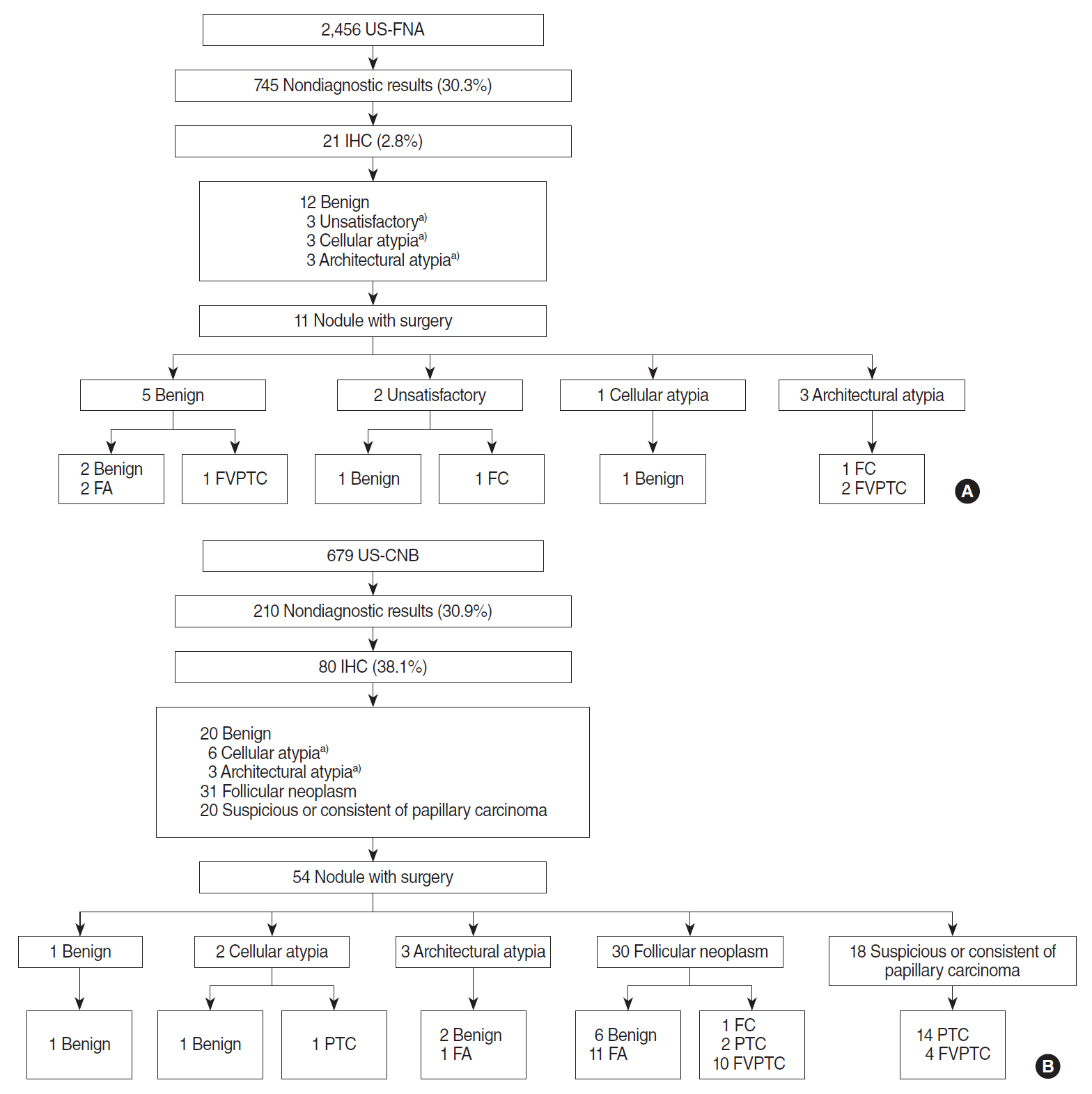
Fig. 1. Flowchart of included cases with immunohistochemical staining (IHC). (A) IHC in fine needle aspiration (FNA) cases. (B) IHC in core needle biopsy (CNB) cases. US-FNA, ultrasound-guided fine needle aspiration; FA, follicular adenoma; FVPTC, follicular variant papillary thyroid carcinoma; FC, follicular carcinoma; US-CNB, ultrasound-guided core needle biopsy; PTC, papillary thyroid carcinoma. a)Indicates cases that still had no diagnosis after IHC staining.

Fig. 2. Heat map of expression of each molecular marker following fine-needle aspiration or core needle biopsy. (A) Expression according to preoperative diagnosis after immunohistochemistry. (B) Expression according to the surgical pathology.
Reference
-
1. Nechifor-Boila A, Catana R, Loghin A, Radu TG, Borda A. Diagnostic value of HBME-1, CD56, Galectin-3 and Cytokeratin-19 in papillary thyroid carcinomas and thyroid tumors of uncertain malignant potential. Rom J Morphol Embryol. 2014; 55(1):49–56.2. Na DG, Kim JH, Sung JY, Baek JH, Jung KC, Lee H, et al. Core-needle biopsy is more useful than repeat fine-needle aspiration in thyroid nodules read as nondiagnostic or atypia of undetermined significance by the Bethesda system for reporting thyroid cytopathology. Thyroid. 2012; May. 22(5):468–75.
Article3. Doddi S, Chohda E, Maghsoudi S, Sheehan L, Sinha A, Chandak P, et al. The final outcome of indeterminate cytology of thyroid nodules in a District General Hospital. G Chir. 2015; May-Jun. 36(3):122–7.
Article4. Na DG, Baek JH, Jung SL, Kim JH, Sung JY, Kim KS, et al. Core needle biopsy of the thyroid: 2016 consensus statement and recommendations from Korean Society of Thyroid Radiology. Korean J Radiol. 2017; Jan-Feb. 18(1):217–37.
Article5. Saleh HA, Jin B, Barnwell J, Alzohaili O. Utility of immunohistochemical markers in differentiating benign from malignant follicular-derived thyroid nodules. Diagn Pathol. 2010; Jan. 5:9.
Article6. Choi YJ, Baek JH, Suh CH, Shim WH, Jeong B, Kim JK, et al. Core-needle biopsy versus repeat fine-needle aspiration for thyroid nodules initially read as atypia/follicular lesion of undetermined significance. Head Neck. 2017; Feb. 39(2):361–9.
Article7. Jung CK, Min HS, Park HJ, Song DE, Kim JH, Park SY, et al. Pathology reporting of thyroid core needle biopsy: a proposal of the Korean Endocrine Pathology Thyroid Core Needle Biopsy Study Group. J Pathol Transl Med. 2015; Jul. 49(4):288–99.
Article8. Ahn SH, Kim SD, Jeong WJ. Comparison of risk of malignancy in a subgroup with atypia of undetermined significance/follicular lesion of undetermined significance: a meta-analysis. Head Neck. 2017; Aug. 39(8):1699–710.
Article9. Kim SD, Han SH, Jeong WJ, Kim H, Ahn SH. Differences in clinical features between subcategories of “atypia/follicular lesion of undetermined significance”. Endocr Pathol. 2017; Sep. 28(3):247–52.
Article10. Li XY, Zhang B, Lin YS. The interpretation of 2015 American Thyroid Association management guidelines for adult patients with thyroid nodules and differentiated thyroid cancer. Zhonghua Er Bi Yan Hou Tou Jing Wai Ke Za Zhi. 2017; Apr. 52(4):309–15.11. Saggiorato E, De Pompa R, Volante M, Cappia S, Arecco F, Dei Tos AP, et al. Characterization of thyroid ‘follicular neoplasms’ in fine-needle aspiration cytological specimens using a panel of immunohistochemical markers: a proposal for clinical application. Endocr Relat Cancer. 2005; Jun. 12(2):305–17.
Article12. Fadda G, Rossi ED, Raffaelli M, Pontecorvi A, Sioletic S, Morassi F, et al. Follicular thyroid neoplasms can be classified as low- and high-risk according to HBME-1 and Galectin-3 expression on liquid-based fine-needle cytology. Eur J Endocrinol. 2011; Sep. 165(3):447–53.
Article13. Trimboli P, Guidobaldi L, Amendola S, Nasrollah N, Romanelli F, Attanasio D, et al. Galectin-3 and HBME-1 improve the accuracy of core biopsy in indeterminate thyroid nodules. Endocrine. 2016; Apr. 52(1):39–45.
Article14. Chiu CG, Strugnell SS, Griffith OL, Jones SJ, Gown AM, Walker B, et al. Diagnostic utility of galectin-3 in thyroid cancer. Am J Pathol. 2010; May. 176(5):2067–81.
Article15. Trimboli P, Nasrollah N, Guidobaldi L, Taccogna S, Cicciarella Modica DD, Amendola S, et al. The use of core needle biopsy as first-line in diagnosis of thyroid nodules reduces false negative and inconclusive data reported by fine-needle aspiration. World J Surg Oncol. 2014; Mar. 12:61.
Article16. Ahn SH, Park SY, Choi SI. Comparison of consecutive results from fine needle aspiration and core needle biopsy in thyroid nodules. Endocr Pathol. 2017; Dec. 28(4):332–8.
Article17. Bartolazzi A, Gasbarri A, Papotti M, Bussolati G, Lucante T, Khan A, et al. Application of an immunodiagnostic method for improving preoperative diagnosis of nodular thyroid lesions. Lancet. 2001; May. 357(9269):1644–50.
Article
- Full Text Links
-
- Actions
-
Cited
- CITED
-
- Close
- Share
-
- Similar articles
-
- Thyroid Nodules with Nondiagnostic FNA Results: Role of Core Needle Biopsy
- Indications for Fine Needle Aspiration in Thyroid Nodules
- Effectiveness and Limitations of Core Needle Biopsy in the Diagnosis of Thyroid Nodules: Review of Current Literature
- Arterial Bleeding of a Thyroid Mass After Thyroid Fine-Needle Aspiration Biopsy: A Case Report
- Natural Course of Cytologically Diagnosed Benign Thyroid Nodules
