Anat Cell Biol.
2018 Sep;51(3):209-211. 10.5115/acb.2018.51.3.209.
Double pouched, sigmoid gallbladder that can cause a diagnostic dilemma to radiologists: a case report
- Affiliations
-
- 1Department of Anatomy, Melaka Manipal Medical College (Manipal Campus), Manipal Academy of Higher Education, Manipal, India. ashwini.anat@gmail.com
- KMID: 2447015
- DOI: http://doi.org/10.5115/acb.2018.51.3.209
Abstract
- Gallbladder shows frequent variations in position, shape, interior, and its duct system. These variations may go unnoticed lifelong; however, they may predispose it for cholecystitis and cholelithiasis. We observed a double pouched gallbladder in an adult male cadaver. The gallbladder was folded to have a sigmoid shape. It had two broad pouches: anterior and posterior and a narrow isthmus in between. Its anterior pouch was covered by peritoneum, whereas the posterior pouch was covered by extrahepatic connective tissue. We discuss the clinical and radiological importance of the case.
Keyword
MeSH Terms
Figure
-

Fig. 1 Photograph of postero-inferior aspect of liver showing the double pouched, folded gallbladder. CL, caudate lobe; GB, gallbladder; IVC, inferior vena cava; LL, left lobe; PFGB, posterior fossa for gallbladder; QL, quadrate lobe.
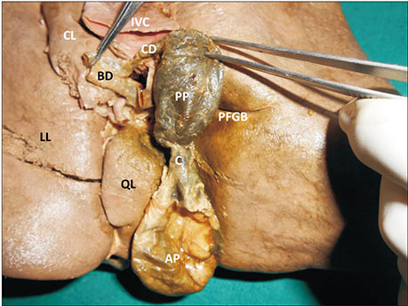
Fig. 2 Photograph of a closer view of the double pouched, folded gallbladder after unfolding it. AP, anterior pouch of gallbladder; BD, bile duct; C, middle constricted part (isthmus); CD, cystic duct; CL, caudate lobe; IVC, inferior vena cava; LL, left lobe; PFGB, posterior fossa for gallbladder; PP, posterior pouch of gallbladder; QL, quadrate lobe.
Reference
-
1. Koszman B. Gall-bladder duplication: case report. Pol Przegl Chir. 2014; 86:433–435.2. Nayak SB, Shetty SD, Surendran S, Jetti R, Kumar N, Sirasanagandla SR. Double gallbladder completely enclosed in a cystogastric fold of peritoneum. Anat Cell Biol. 2014; 47:132–134.
Article3. Esper E, Kaufman DB, Crary GS, Snover DC, Leonard AS. Septate gallbladder with cholelithiasis: a cause of chronic abdominal pain in a 6-year-old child. J Pediatr Surg. 1992; 27:1560–1562.
Article4. van Kamp MJ, Bouman DE, Steenvoorde P, Klaase JM. A phrygian cap. Case Rep Gastroenterol. 2013; 7:347–351.
Article5. Hibbs H, Ahmad U. Inverted liver with suprahepatic, anteriorly displaced gallbladder. J La State Med Soc. 2010; 162:150–152.6. Ishii H, Noguchi A, Onishi M, Takao K, Maruyama T, Taiyoh H, Araki Y, Shimizu T, Izumi H, Tani N, Yamaguchi M, Yamane T. True left-sided gallbladder with variations of bile duct and cholecystic vein. World J Gastroenterol. 2015; 21:6754–6758.
Article7. Miyakura Y, Sadatomo A, Ohta M, Lefor AT, Sata N, Nishimura N, Sakatani T, Yasuda Y. Floating gallbladder strangulation caused by the lesser omentum: report of a case. Surg Today. 2012; 42:693–696.
Article8. Pillay Y. Gallbladder duplication. Int J Surg Case Rep. 2015; 11:18–20.
Article
- Full Text Links
-
- Actions
-
Cited
- CITED
-
- Close
- Share
-
- Similar articles
-
- Double Primary Cancer of the Gallbladder and Cystic Duct: A Case Report
- A Case af Chronic Cholecystitis Diagnosed with Double Contrast Study of the Gallbladder
- A Case of a Double Gallbladder with Common Bile Duct Stones
- Two Cases of Gallbladder Diseases Diagnosed with Double Contrast Study of the Gallbladder
- A Case of Synchronous Double Primary Cancer Associated with the Biliary Tract
