Cauda Equine Syndrome Induced by Spinal Epidural Hematoma due to a Low-Energy Osteoporotic Vertebral Fracture
- Affiliations
-
- 1Department of Orthopedic Surgery, Sung-Ae Hospital, Seoul, Korea. hsh315@naver.com
- KMID: 2444786
- DOI: http://doi.org/10.4055/jkoa.2019.54.2.187
Abstract
- Spinal epidural hematoma (SEH) can occur naturally or traumatically and is most common in patients with an underlying disease of the vascular structure or coagulation disorder. Most SEHs occur naturally for no apparent reason, and epidural hematoma caused by trauma is less common, comprising 1.0%-1.7% of total spinal injuries. Few reports of SEH induced cauda equine syndrome resulting from low-energy injury caused by osteoporotic vertebral compression fractures are available. The authors experienced a case of delayed SEH after hemorrhage due to a low-energy injury in an elderly patient. No cases in Korea have been reported; therefore, this case is reported with a review of the relevant literature.
Keyword
MeSH Terms
Figure
-
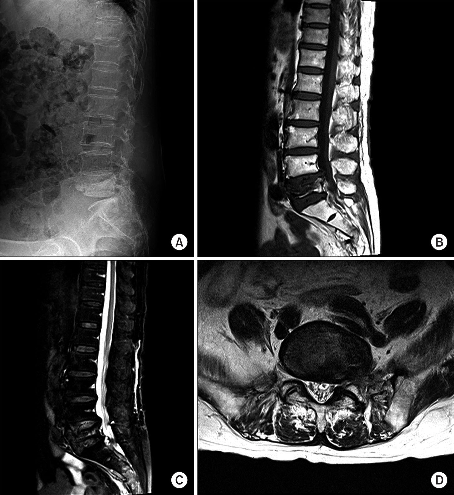
Figure 1. Initial lateral Radiography (A), T1 weighted sagittal magnetic resonance image (B), and T2 weighted fat suppression sagittal magnetic resonance image (C) demonstrating a recent compression fracture of the L5 vertebral body compression rate 30.2%. T2 weighted axial magnetic resonance image through the L5 vertebra (D) showing no visible epidural hematoma.
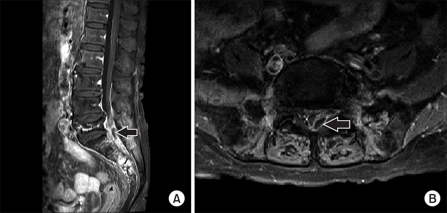
Figure 2. Preoperation magnetic resonance imaging studies demonstrating epidural hematoma. (A) Gadolinium enhanced T1 weighted sagittal magnetic resonance image shows epidural lesion (hematoma) (arrow) showing intermediate high signal, compressing dural sac. (B) Gadolinium enhanced T1 weighted axial magnetic resonance image shows that the left posterolateral dural sac (arrow) is a compressed epidural lesion at L5-S1.
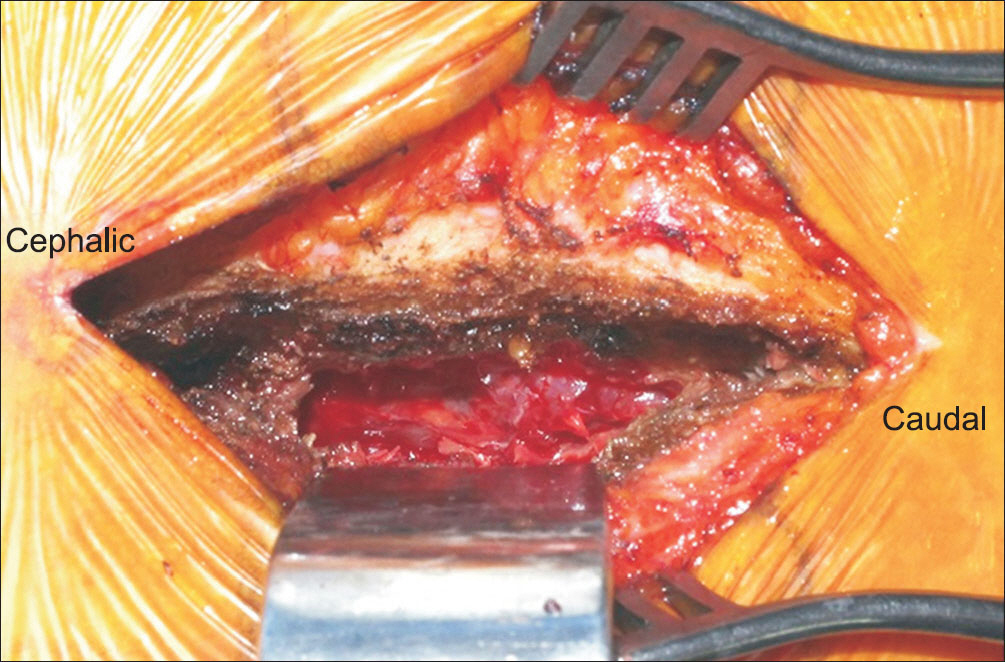
Figure 3. Intraoperative photograph shows that the left S1 root was compressed due to an epidural hematoma.
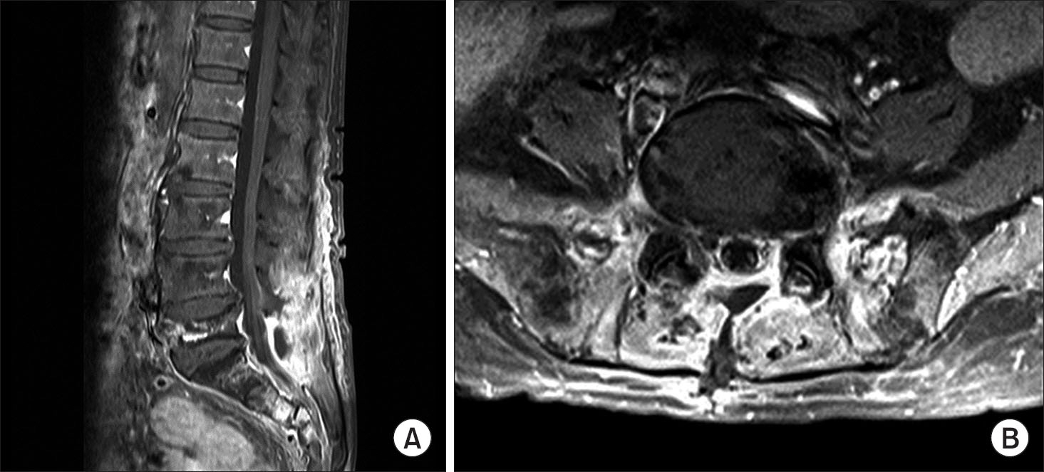
Figure 4. One months later, gadolinium enhanced T1 weighted sagittal magnetic resonance image (A) and gadolinium enhanced T1 weighted axial magnetic resonance image (B) showed no more visible nonenhanced left posterolateral epidural lesion at L5-S1.
Reference
-
References
1. Rechtine GR 2nd, Bolesta MJ, Chrin AM, Louis K. Spontaneous resolution of symptomatic post-traumatic cervical epidural hematoma. J Bone Joint Surg Am. 2001. 83-A:255–8.
Article2. Hsieh CT, Chiang YH, Tang CT, Sun JM, Ju DT. Delayed traumatic thoracic spinal epidural hematoma: a case report and literature review. Am J Emerg Med. 2007. 25:69–71.
Article3. Cuenca PJ, Tulley EB, Devita D, Stone A. Delayed traumatic spinal epidural hematoma with spontaneous resolution of symptoms. J Emerg Med. 2004. 27:37–41.
Article4. Kreppel D, Antoniadis G, Seeling W. Spinal hematoma: a literature survey with meta-analysis of 613 patients. Neurosurg Rev. 2003. 26:1–49.
Article5. Rodrigues LM, Abreu F, Fujiki EN, Milani C. Delayed traumatic spinal epidural hematoma with neurological deficits. Einstein (Sao Paulo). 2010. 8:477–9.
Article6. Adhiyaman V, Asghar M, Ganeshram KN, Bhowmick BK. Chronic subdural haematoma in the elderly. Postgrad Med J. 2002. 78:71–5.7. Boukobza M, Guichard JP, Boissonet M. . Spinal epidural haematoma: report of 11 cases and review of the literature. Neuroradiology. 1994. 36:456–9.
Article8. Jamjoom ZA. Acute spontaneous spinal epidural hematoma: the influence of magnetic resonance imaging on diagnosis and treatment. Surg Neurol. 1996. 46:345–9.
Article9. Lawton MT, Porter RW, Heiserman JE, Jacobowitz R, Sonntag VK, Dickman CA. Surgical management of spinal epidural hematoma: relationship between surgical timing and neurological outcome. J Neurosurg. 1995. 83:1–7.
Article
- Full Text Links
-
- Actions
-
Cited
- CITED
-
- Close
- Share
-
- Similar articles
-
- Thoracolumbar Epidural Hematoma Complicated by Cauda Equina Syndrome : Complication of Systemic Heparinization Following Epidural Anesthesia: A case report
- Delayed Spinal Epidural Hematoma after En Block Spondylectomy for Vertebral Ewing's Sarcoma
- Cauda Equina Syndrome from Subdural Hematoma after Caudal Epidural Injection
- Lumbar Spinal Epidural Abscess Combined with Cauda Equina Syndrome: A Case Report
- Cauda Equina Syndrome due to Epidural Hematoma After Lumbar Epidural Block : A Case Report
