Atlanto-Occipital Dislocation: A Case Report
- Affiliations
-
- 1Department of Neurosurgery, Dongguk University Gyeongju Hospital, Dongguk University College of Medicine, Gyeongju, Korea. doctorkim128@naver.com
- KMID: 2444207
- DOI: http://doi.org/10.13004/kjnt.2019.15.e3
Abstract
- Patients with atlanto-occipital dislocation (AOD) are increasingly being transported to emergency rooms, alive, by the improved pre-hospital emergency rescue system. The author reports a fatal case of AOD with severe neurovascular injuries following a high-speed pedestrian collision. Therefore, nowadays, neurosurgeons can expect an increase in the occurrence of such cases; an early diagnosis and prompt occipitocervical fusion can save lives. This report reviews the current concepts of AOD in mild to fatal conditions.
MeSH Terms
Figure
-
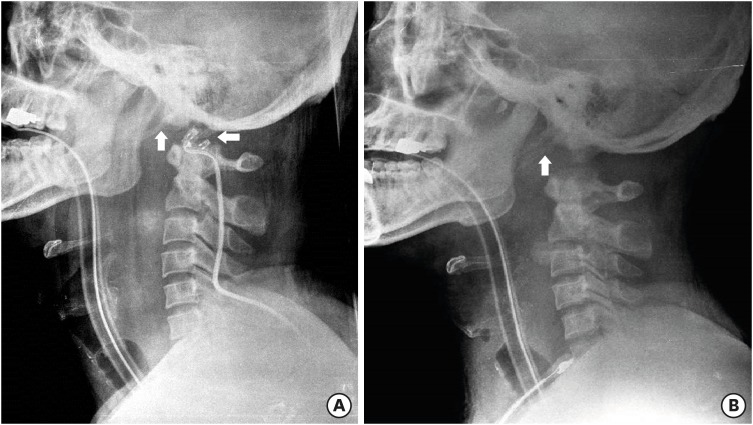
FIGURE 1 Initial (A) and 8 hours follow-up (B) cervical X-ray lateral views show atlanto-occipital dislocation and instability. These images (A, B) show mixed type I and II according to Traynelis et al.20)
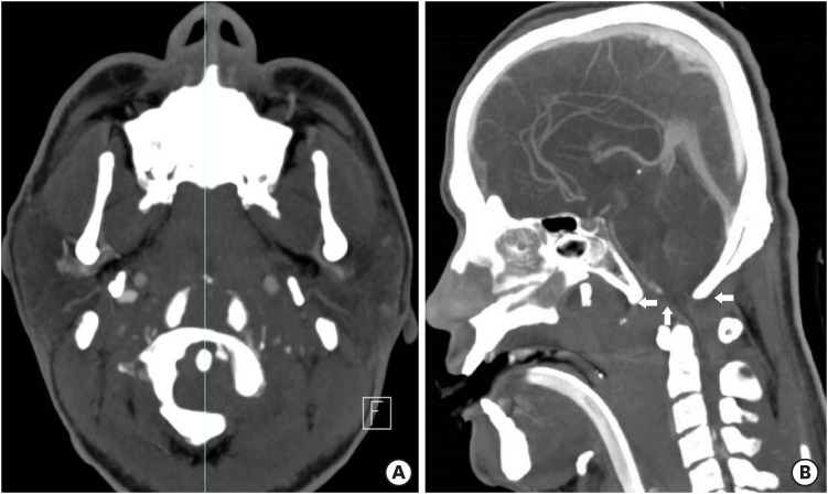
FIGURE 2 Three dimensional computed tomography angiography of axial (C1) and midsagittal reconstruction view show atlanto-occipital dislocation and associated lesion of medulla oblongata and upper cervical cord. (A) Axial view shows double contour of occipital condyles in front of anterior arch of atlas. Axial view shows the level of midsagittal view (B). (B) Midsagittal view shows the huge hematoma at retropharyngeal and retropharyngeal space, around medullar oblongata and upper cervical cord, thinning of medulla and anterior dislocation of foramen magnum.
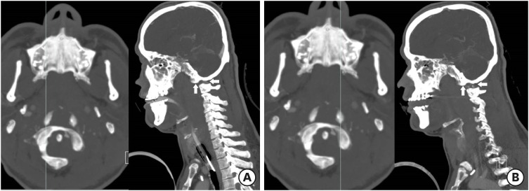
FIGURE 3 Sagittally reformatted computed tomography images show dislocation of the right (A) and left (B) atlanto-occipital joints.
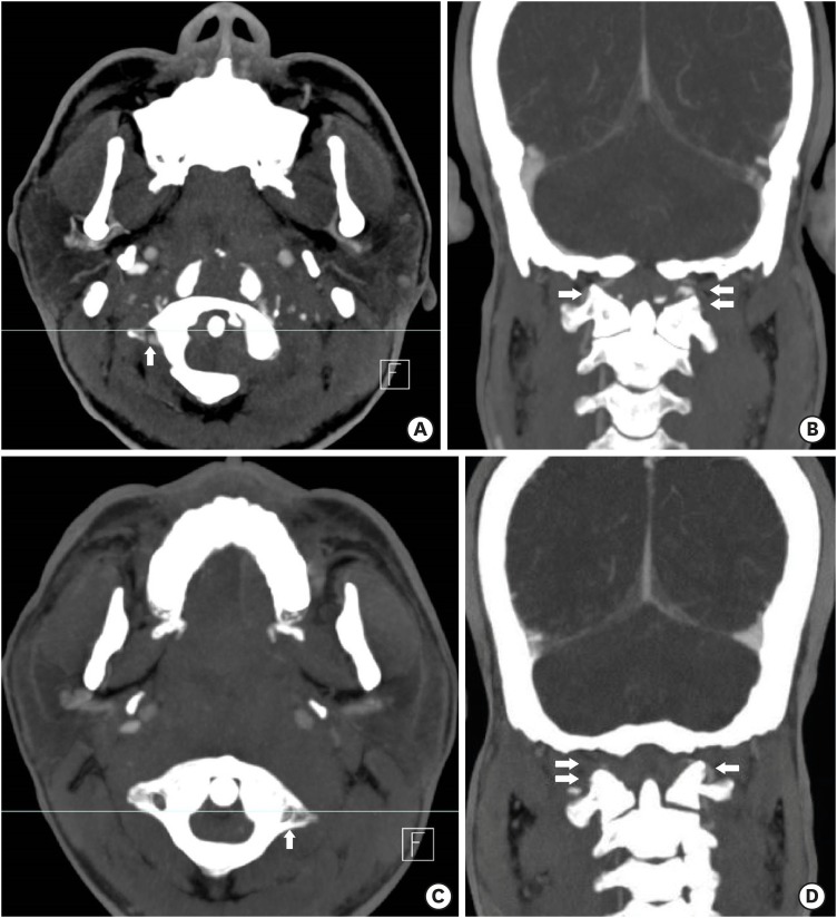
FIGURE 4 Three dimensional computed tomography angiography of axial (C1) and sagittal reconstruction views show atlanto-occipital dislocation and associated lesion of left vertebral artery at C1 level. (A) Axial view showing the level of coronal section (B) at right transverse foramen of atalas. Axial view shows double contour of occipital condyles in front of anterior arch of atlas due to anterior dislocation. (B) Coronal reconstruction view at right transverse foramen of atlas shows the continuity of right vertebral artery at C1 level. (C) Axial view showing the level of coronal section (D) at left transverse foramen of atalas. (D) Coronal reconstruction view at left transverse foramen of atlas shows the loss of continuity of left vertebral artery at C1 level revealing dissection of left vertebral artery.
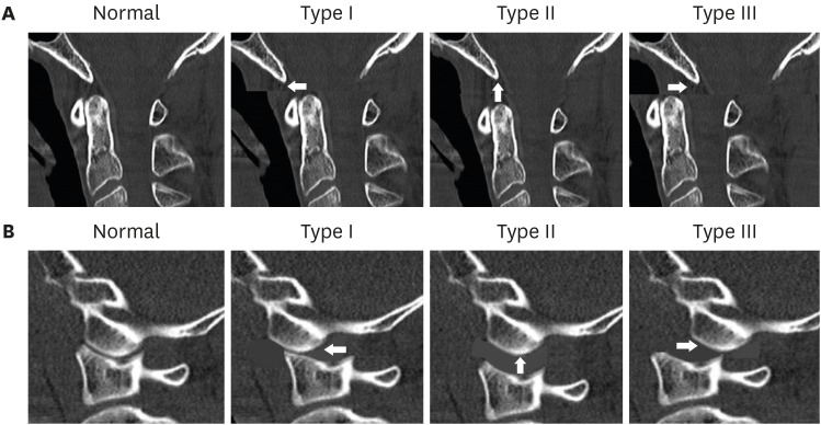
FIGURE 5 The Traynelis et al.20) classification. type I (anterior dislocation), type II (longitudinal separation), and type III (posterior dislocation). (A) Midsagittal plane views show the relation of the basion, the posterior arch of C1, the opisthion and the anterior arch of the atlas. (B) Parasagittal plane views show the relation of the occipital condyle and the superior articular surface of lateral mass for occipital condyle.
Reference
-
1. Alker GJ, Oh YS, Leslie EV, Lehotay J, Panaro VA, Eschner EG. Postmortem radiology of head neck injuries in fatal traffic accidents. Radiology. 1975; 114:611–617. PMID: 1118566.2. Astur N, Klimo P Jr, Sawyer JR, Kelly DM, Muhlbauer MS, Warner WC Jr. Traumatic atlanto-occipital dislocation in children: evaluation, treatment, and outcomes. J Bone Joint Surg Am. 2013; 95:e194. PMID: 24352780.3. Blackwood NJ. III. Atlo-occipital dislocation: a case of fracture of the atlas and axis, and forward dislocation of the occiput on the spinal column, life being maintained for thirty-four hours and forty minutes by artificial respiration, during which a laminectomy was performed upon the third cervical vertebra. Ann Surg. 1908; 47:654–658. PMID: 17862147.4. Dahdaleh NS, Khanna R, Menezes AH, Smith ZA, Viljoen SV, Koski TR, et al. The application of the revised condyle-C1 interval method to diagnose traumatic atlanto-occipital dissociation in adults. Global Spine J. 2016; 6:529–534. PMID: 27555993.
Article5. Garrett M, Consiglieri G, Kakarla UK, Chang SW, Dickman CA. Occipitoatlantal dislocation. Neurosurgery. 2010; 66:48–55.
Article6. Hall GC, Kinsman MJ, Nazar RG, Hruska RT, Mansfield KJ, Boakye M, et al. Atlanto-occipital dislocation. World J Orthop. 2015; 6:236–243. PMID: 25793163.
Article7. Harris JH Jr, Carson GC, Wagner LK. Radiologic diagnosis of traumatic occipitovertebral dissociation: 1. Normal occipitovertebral relationships on lateral radiographs of supine subjects. AJR Am J Roentgenol. 1994; 162:881–886. PMID: 8141012.
Article8. Harris JH Jr, Carson GC, Wagner LK, Kerr N. Radiologic diagnosis of traumatic occipitovertebral dissociation: 2. Comparison of three methods of detecting occipitovertebral relationships on lateral radiographs of supine subjects. AJR Am J Roentgenol. 1994; 162:887–892. PMID: 8141013.
Article9. Hauswald M, Sklar DP, Tandberg D, Garcia JF. Cervical spine movement during airway management: cinefluoroscopic appraisal in human cadavers. Am J Emerg Med. 1991; 9:535–538. PMID: 1930391.
Article10. Jeszenszky D, Fekete TF, Lattig F, Bognár L. Intraarticular atlantooccipital fusion for the treatment of traumatic occipitocervical dislocation in a child: a new technique for selective stabilization with nine years follow-up. Spine (Phila Pa 1976). 2010; 35:E421–E426. PMID: 20393390.11. Kim YJ, Yoo CJ, Park CW, Lee SG, Son S, Kim WK. Traumatic atlanto-occipital dislocation (AOD). Korean J Spine. 2012; 9:85–91. PMID: 25983794.
Article12. Lee C, Woodring JH, Goldstein SJ, Daniel TL, Young AB, Tibbs PA. Evaluation of traumatic atlantooccipital dislocations. AJNR Am J Neuroradiol. 1987; 8:19–26. PMID: 3101469.13. Pang D, Nemzek WR, Zovickian J. Atlanto-occipital dislocation: part 1--normal occipital condyle-C1 interval in 89 children. Neurosurgery. 2007; 61:514–521. PMID: 17881963.14. Pang D, Nemzek WR, Zovickian J. Atlanto-occipital dislocation--part 2: the clinical use of (occipital) condyle-C1 interval, comparison with other diagnostic methods, and the manifestation, management, and outcome of atlanto-occipital dislocation in children. Neurosurgery. 2007; 61:995–1015. PMID: 18091277.15. Park MS, Moon SH, Kim TH, Oh JK, Nam JH, Jung JK, et al. New radiographic index for occipito-cervical instability. Asian Spine J. 2016; 10:123–128. PMID: 26949467.
Article16. Payer M, Sottas CC. Traumatic atlanto-occipital dislocation: presentation of a new posterior occipitoatlantoaxial fixation technique in an adult survivor: technical case report. Neurosurgery. 2005; 56:E203. PMID: 15799814.
Article17. Powers B, Miller MD, Kramer RS, Martinez S, Gehweiler JA Jr. Traumatic anterior atlanto-occipital dislocation. Neurosurgery. 1979; 4:12–17. PMID: 450210.
Article18. Smith KM, Yoganandan N, Pintar FA, Kurpad SN, Maiman DJ. Atlantooccipital dislocation in motor vehicle side impact, derivation of the mechanism of injury, and implications for early diagnosis. J Craniovertebr Junction Spine. 2010; 1:113–117. PMID: 21572632.19. Theodore N, Aarabi B, Dhall SS, Gelb DE, Hurlbert RJ, Rozzelle CJ, et al. The diagnosis and management of traumatic atlanto-occipital dislocation injuries. Neurosurgery. 2013; 72(Suppl 2):114–126. PMID: 23417184.
Article20. Traynelis VC, Marano GD, Dunker RO, Kaufman HH. Traumatic atlanto-occipital dislocation. Case report. J Neurosurg. 1986; 65:863–870. PMID: 3772485.
- Full Text Links
-
- Actions
-
Cited
- CITED
-
- Close
- Share
-
- Similar articles
-
- Traumatic Atlanto-Occipital Rotatory Posterior Dislocation Combined with Atlanto-Axial Rotatory Subluxation: A Case Report
- Traumatic Atlanto-Occipital Dislocation: A Case Report
- Atlanto-occipital Assimilation Can be Misdiagnosed as Atlantoaxial Dislocation: A Case Report
- Traumatic Atlanto-Occipital Dislocation Presenting With Dysphagia as the Chief Complaint: A Case Report
- A Case of Congenital Atlanto-Occipital Fusion: One case report
