Pseudo-Subarachnoid Hemorrhage; Chronic Subdural Hematoma with an Unruptured Aneurysm Mistaken for Subarachnoid Hemorrhage
- Affiliations
-
- 1Department of Neurosurgery, Pusan National University Yangsan Hospital, Yangsan, Korea. twins728@hanmail.net
- KMID: 2444202
- DOI: http://doi.org/10.13004/kjnt.2019.15.e11
Abstract
- Subarachnoid hemorrhage (SAH) usually occurs due to aneurysmal rupture of intracranial arteries and its typical computed tomography (CT) findings are increased attenuation of cisterns and subarachnoid spaces. However, several CT findings mimicking SAH are feasible in diverse conditions. They are so-called as pseudo-SAH, and this report is a case of pseudo-SAH which is misdiagnosed as aneurysm rupture accompanied by bilateral chronic subdural hematoma (cSDH). A 42-year-old male with severe headache visited our institute. Non-contrast brain CT images showed increased attenuation on basal cistern, and cSDH on both fronto-temporo-parietal convexity with midline shifting. Trans-femoral cerebral angiography was done and we confirmed small aneurysm at right M1 portion of middle cerebral artery. Under diagnosis of SAH, we planned an operation in order to clip aneurysmal neck and remove cSDH. cSDH was removed as planned, however, there was no SAH and we also couldn't find the rupture point of aneurysm. Serial follow-up CT showed mild cumulative cSDH recurrence, but the patient was tolerant and had no neurologic deficit during hospitalization. We have checked the patient via out-patient department for 6 months, there are no significant changes in volume and density of cSDH and the patient also have no neurologic complications.
Keyword
MeSH Terms
-
Adult
Aneurysm*
Arteries
Brain
Brain Edema
Cerebral Angiography
Diagnosis
Follow-Up Studies
Headache
Hematoma, Subdural, Chronic*
Hemorrhage*
Hospitalization
Humans
Intracranial Hypertension
Male
Middle Cerebral Artery
Neck
Neurologic Manifestations
Outpatients
Recurrence
Rupture
Subarachnoid Hemorrhage*
Subarachnoid Space
Figure
-
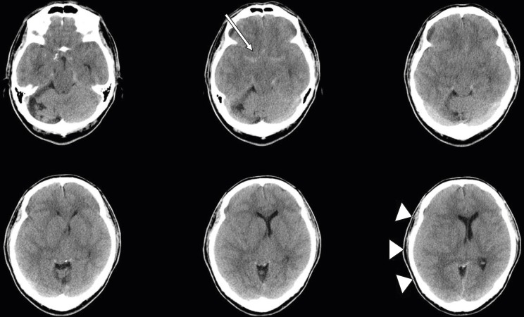
FIGURE 1 Initial non-contrast brain computed tomography findings showed increased attenuation at basal cistern (arrow) and fluid collection along the right fronto-parietal convexity (triangles) with midline shifting.
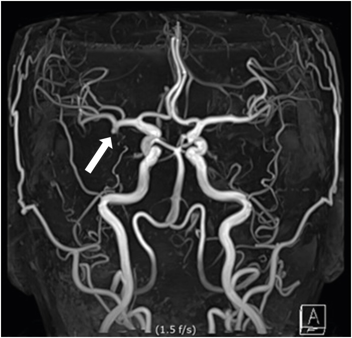
FIGURE 2 Previous magnetic resonance angiography showed unruptured aneurysm at right M1 (arrow).
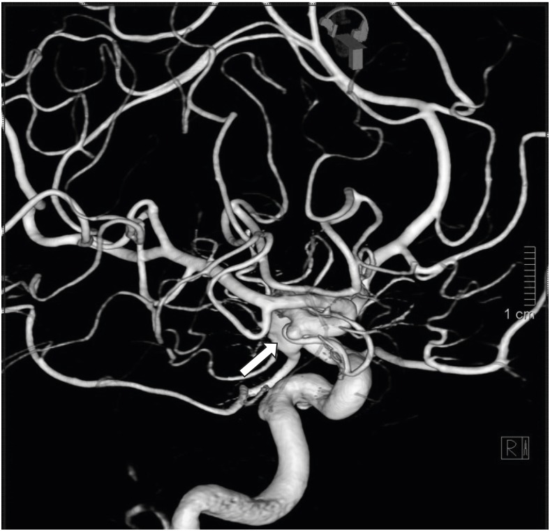
FIGURE 3 Trans-femoral cerebral angiography was performed, and then small aneurysm at right anterior temporal branch origin of middle cerebral artery M1 portion was confirmed (arrow).
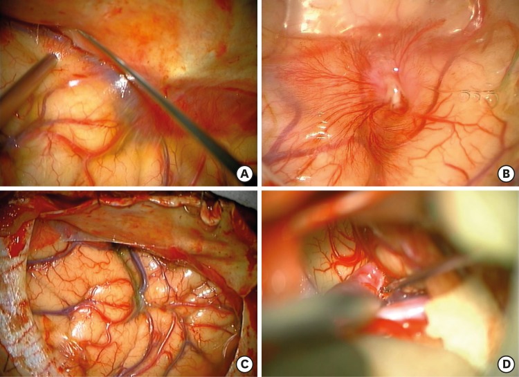
FIGURE 4 (A) Vascularized inner membrane of cSDH looks like arachnoid adhesion. (B) Neovascularization of inner membrane of cSDH was also found. (C) After resection of inner membrane of cSDH, normal arachnoid membrane was observed. (D) No subarachnoid hemorrhage was observed during sylvian fissure dissection.cSDH: chronic subdural hematoma.
Reference
-
1. al-Yamany M, Deck J, Bernstein M. Pseudo-subarachnoid hemorrhage: a rare neuroimaging pitfall. Can J Neurol Sci. 1999; 26:57–59. PMID: 10068810.2. Ammerman JM. Pseudosubarachnoid hemorrhage: a zebra worth looking for. South Med J. 2008; 101:1200. PMID: 19005457.
Article3. Coady G, Brewer M, Maillloux P. Pseudosubarachnoid hemorrhage in a 42-year-old male with meningitis. Radiol Case Rep. 2015; 6:470. PMID: 27307899.
Article4. Ducruet AF, Grobelny BT, Zacharia BE, Hickman ZL, DeRosa PL, Andersen KN, et al. The surgical management of chronic subdural hematoma. Neurosurg Rev. 2012; 35:155–169. PMID: 21909694.
Article5. Eckel TS, Breiter SN, Monsein LH. Subarachnoid contrast enhancement after spinal angiography mimicking diffuse subarachnoid hemorrhage. AJR Am J Roentgenol. 1998; 170:503–505. PMID: 9456974.
Article6. Ferrante E, Regna-Gladin C, Arpino I, Rubino F, Porrinis L, Ferrante MM, et al. Pseudo-subarachnoid hemorrhage: a potential imaging pitfall associated with spontaneous intracranial hypotension. Clin Neurol Neurosurg. 2013; 115:2324–2328. PMID: 24075686.
Article7. Given CA 2nd, Burdette JH, Elster AD, Williams DW 3rd. Pseudo-subarachnoid hemorrhage: a potential imaging pitfall associated with diffuse cerebral edema. AJNR Am J Neuroradiol. 2003; 24:254–256. PMID: 12591643.8. Hasan TF, Duarte W, Akinduro OO, Goldstein ED, Hurst R, Haranhalli N, et al. Nonaneurysmal “pseudo-subarachnoid hemorrhage” computed tomography patterns: Challenges in an acute decision-making heuristics. J Stroke Cerebrovasc Dis. 2018; 27:2319–2326. PMID: 29884521.
Article9. Isobe N, Sato H, Murakami T, Kurokawa Y, Seyama G, Oki S. Six cases of organized chronic subdural hematoma. No Shinkei Geka. 2008; 36:1115–1120. PMID: 19086442.10. Lin CY, Lai PH, Fu JH, Wang PC, Pan HB. Pseudo-subarachnoid hemorrhage: a potential imaging pitfall. Can Assoc Radiol J. 2014; 65:225–231. PMID: 24325921.
Article11. Mendelsohn DB, Moss ML, Chason DP, Muphree S, Casey S. Acute purulent leptomeningitis mimicking subarachnoid hemorrhage on CT. J Comput Assist Tomogr. 1994; 18:126–128. PMID: 8282861.
Article12. Noguchi K, Ogawa T, Inugami A, Toyoshima H, Sugawara S, Hatazawa J, et al. Acute subarachnoid hemorrhage: MR imaging with fluid-attenuated inversion recovery pulse sequences. Radiology. 1995; 196:773–777. PMID: 7644642.
Article13. Rabinstein AA, Pittock SJ, Miller GM, Schindler JJ, Wijdicks EF. Pseudosubarachnoid haemorrhage in subdural haematoma. J Neurol Neurosurg Psychiatry. 2003; 74:1131–1132. PMID: 12876252.
Article14. Rocchi G, Caroli E, Salvati M, Delfini R. Membranectomy in organized chronic subdural hematomas: indications and technical notes. Surg Neurol. 2007; 67:374–380. PMID: 17350406.
Article15. Senthilkumaran S, Balamurugan N, Menezes RG, Thirumalaikolundusubramanian P. Role of Hounsfield units to distinguish pseudo-subarachnoid hemorrhage. Clin Toxicol (Phila). 2011; 49:948. PMID: 22070557.
Article16. Senthilkumaran S, Sweni S, Balamurugan N, Jena NN, Thirumalaikolundusubramanian P. Hounsfield units in pseudosubarachnoid hemorrhage-an old yet fascinating tool. Am J Emerg Med. 2015; 33:1095.
Article17. Spiegel SM, Fox AJ, Vinuela F, Pelz DM. Increased density of tentorium and falx: a false positive CT sign of subarachnoid hemorrhage. Can Assoc Radiol J. 1986; 37:243–247. PMID: 2950107.
- Full Text Links
-
- Actions
-
Cited
- CITED
-
- Close
- Share
-
- Similar articles
-
- Pure Acute Subdural Hematoma without Subarachnoid Hemorrhage Secondary to Posterior Communicating Artery Aneurysm Rupture: A Case Report
- Spontaneous Spinal Subdural and Subarachnoid Hemorrhage with Concomitant Intracerebral Hemorrhage: A Case Report
- Spontaneous Spinal Subdural Hematoma with Simultaneous Cranial Subarachnoid Hemorrhage
- Pure Subdural Hemorrhage Caused by Internal Carotid Artery Dorsal Wall Aneurysm Rupture
- Subdural Hematoma Due to Ruptured Intracerebral Aneurysm
