Nicorandil alleviated cardiac hypoxia/reoxygenation-induced cytotoxicity via upregulating ketone body metabolism and ACAT1 activity
- Affiliations
-
- 1Department of Cardiology, The Affiliated Hospital of Yan'an University, Yan'an 716000, China.
- 2Department of Cardiology, The Forth Renmin Hospital of Xi'an, Shanxi Province 710004, China. hanleisen196601@126.com
- KMID: 2443614
- DOI: http://doi.org/10.4196/kjpp.2019.23.1.37
Abstract
- To study the effect of nicorandil pretreatment on ketone body metabolism and Acetyl-CoA acetyltransferase (ACAT1) activity in hypoxia/reoxygenation (H/R)-induced cardiomyocytes. In our study, we applied H9c2 cardiomyocytes cell line to evaluate the cardioprotective effects of nicorandil. We detected mitochondrial viability, cellular apoptosis, reactive oxygen species (ROS) production and calcium overloading in H9c2 cells that exposed to H/R-induced cytotoxicity. Then we evaluated whether nicorandil possibly regulated ketone body, mainly β-hydroxybutyrate (BHB) and acetoacetate (ACAC), metabolism by regulating ACAT1 and Succinyl-CoA:3-keto-acid coenzyme A transferase 1 (OXCT1) protein and gene expressions. Nicorandil protected H9c2 cardiomyocytes against H/R-induced cytotoxicity dose-dependently by mitochondria-mediated anti-apoptosis pathway. Nicorandil significantly decreased cellular apoptotic rate and enhanced the ratio of Bcl-2/Bax expressions. Further, nicorandil decreased the production of ROS and alleviated calcium overloading in H/R-induced H9c2 cells. In crucial, nicorandil upregulated ACAT1 and OXCT1 protein expressions and either of their gene expressions, contributing to increased production of cellular BHB and ACAC. Nicorandil alleviated cardiomyocytes H/R-induced cytotoxicity through upregulating ACAT1/OXCT1 activity and ketone body metabolism, which might be a potential mechanism for emerging study of nicorandil and other K(ATP) channel openers.
MeSH Terms
Figure
-
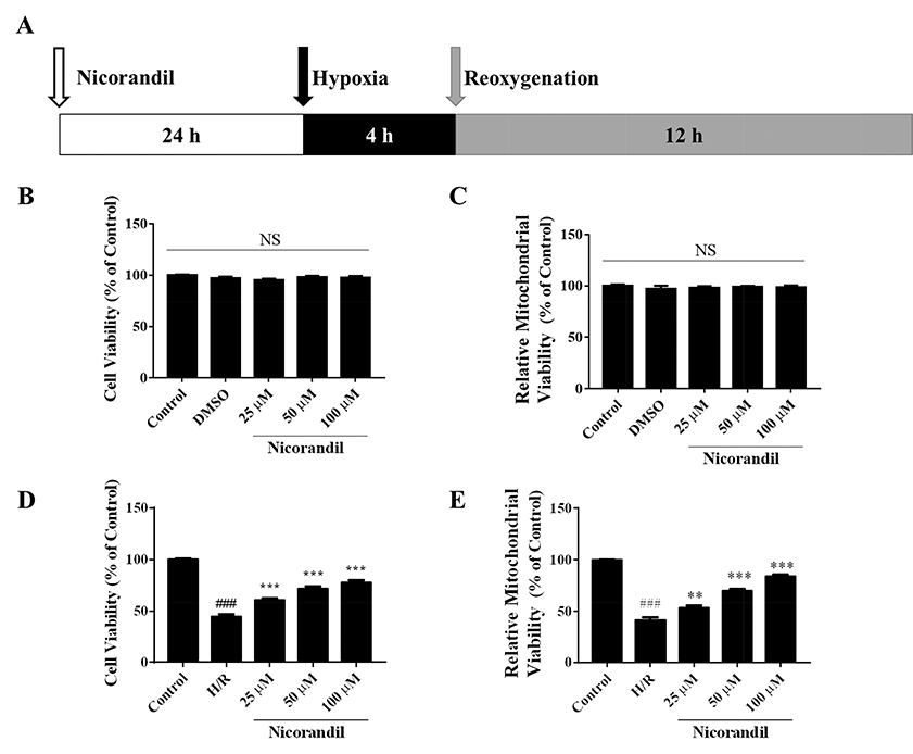
Fig. 1 Effects of nicorandil on cell and mitochondrial viability in H/R-induced H9c2 cells. (A) Hypoxia/reoxygenation protocol and drug treatment. Nicorandil was pretreatment 24 h before H/R. H/R was induced by incubating cells in KRB buffer as hypoxia for 4 h followed by incubating cells in cultural medium as reoxygenation for 12 h. (B, D) Effects of Nicorandil (25, 50, 100 µM) on cell viability under normoxia or hypoxia conditioning determined by MTT assay. Data (n=3) were shown as the mean±SEM, NS: no significance. (C, E) Effects of nicorandil (25, 50, 100 µM) on mitochondrial viability under normoxia or hypoxia conditioning determined by mitochondrial viability assay. Data (n=3) were shown as the mean±SEM, ###p<0.001 vs. control group, **p<0.01 vs. H/R group, ***p<0.001 vs. H/R group.

Fig. 2 Effects of nicorandil on cellular apoptosis, ROS production and calcium overloading in H/R-induced H9c2 cells. (A, B) Effects of nicorandil (25, 50, 100 µM) on cellular apoptosis was detected by PI and Annexin V double staining evaluated by flow cytometry. Data (n=3) were presented as mean±SEM. ###p<0.001 vs. control group, **p<0.01 vs. H/R group, ***p<0.001 vs. H/R group. (C) ROS production was detected by H2DCFDA probe staining and evaluated by flow cytometry. Data (n=3) were presented as mean±SEM. ###p<0.001 vs. control group, **p<0.01 vs. H/R group, ***p<0.001 vs. H/R group. (D) Calcium concentration was detected by Fluo-4 AM staining and evaluated by flow cytometry. Data (n=3) were presented as mean±SEM. ###p<0.001 vs. control group, *p<0.05 vs. H/R group, ***p<0.001 vs. H/R group. (E, F) The expression of Bcl-2 and Bax were detected by western blotting. β-Actin as a loading control. Data (n=3) were presented as mean±SEM. ###p<0.001 vs. control group, *p<0.05 vs. H/R group, ***p<0.001 vs. H/R group.
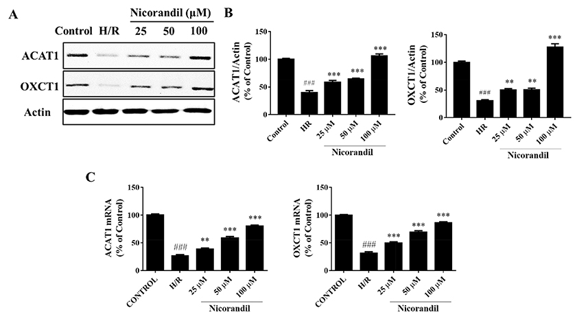
Fig. 3 The effects of nicorandil on ACAT1 and OXCT1 protein expressions in H/R-induced H9c2 cells. (A) The expression of ACAT1 and OXCT1 were detected by western blotting. β-Actin as a loading control. (B) The relative protein expressions were calculated by normalizing to β-Actin. Data (n=3) were presented as mean±SEM. ###p<0.001 vs. control group, **p<0.01 vs. H/R group, ***p<0.001 vs. H/R group. (C) mRNA levels were quantified by real-time RT-PCR. Data (n=3) were presented as mean±SEM. ###p<0.001 vs. control group, **p<0.01 vs. H/R group, ***p<0.001 vs. H/R group.

Fig. 4 Effects of nicorandil on ketone bodies productions in H/R-induced H9c2 cells. (A) The concentration of β-hydroxybutyrate (BHB) in cell extractions. Data (n=3) were presented as mean±SEM. ###p<0.001 vs. control group, **p<0.01 vs. H/R group, ***p<0.001 vs. H/R group. (B) The concentration of acetoacetate (ACAC) in cell extractions. Data (n=3) were presented as mean±SEM. ###p<0.001 vs. control group, ***p<0.001 vs. H/R group.
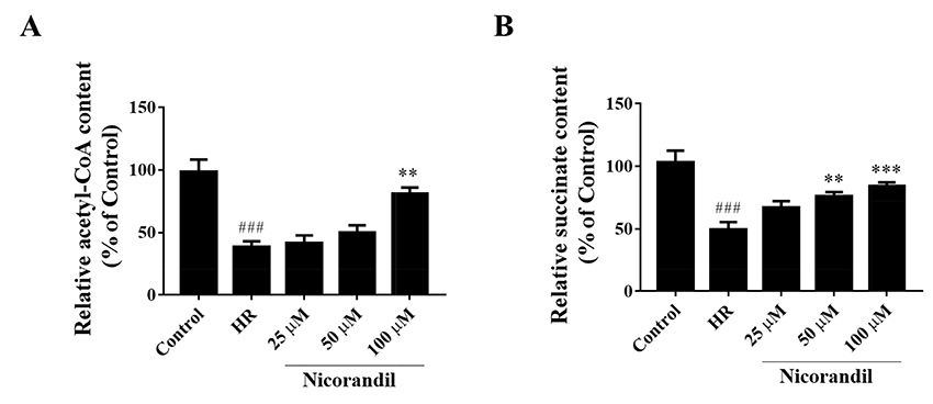
Fig. 5 Effects of nicorandil on productions of acetyl-CoA and succinate in H/R-induced H9c2 cells. (A) The concentration of acetyl-CoA in cell extractions. Data (n=3) were presented as mean±SEM. ###p<0.001 vs. control group, **p<0.01 vs. H/R group. (B) The concentration of succinate in cell extractions. Data (n=3) were presented as mean±SEM. ###p<0.001 vs. control group, **p<0.01 vs. H/R group, ***p<0.001 vs. H/R group.
Reference
-
1. Yellon DM, Hausenloy DJ. Myocardial reperfusion injury. N Engl J Med. 2007; 357:1121–1135.
Article2. Silachev DN, Plotnikov EY, Pevzner IB, Zorova LD, Babenko VA, Zorov SD, Popkov VA, Jankauskas SS, Zinchenko VP, Sukhikh GT, Zorov DB. The mitochondrion as a key regulator of ischaemic tolerance and injury. Heart Lung Circ. 2014; 23:897–904.
Article3. Abete P, Testa G, Cacciatore F, Della-Morte D, Galizia G, Langellotto A, Rengo F. Ischemic preconditioning in the younger and aged heart. Aging Dis. 2011; 2:138–148.4. Kloner RA, Rezkalla SH. Preconditioning, postconditioning and their application to clinical cardiology. Cardiovasc Res. 2006; 70:297–307.
Article5. Fukao T, Mitchell G, Sass JO, Hori T, Orii K, Aoyama Y. Ketone body metabolism and its defects. J Inherit Metab Dis. 2014; 37:541–551.
Article6. Cotter DG, Schugar RC, Crawford PA. Ketone body metabolism and cardiovascular disease. Am J Physiol Heart Circ Physiol. 2013; 304:H1060–H1076.
Article7. Faria MH, Muniz LR, Vasconcelos PR. Ketone bodies metabolism during ischemic and reperfusion brain injuries following bilateral occlusion of common carotid arteries in rats. Acta Cir Bras. 2007; 22:125–129.
Article8. Tieu K, Perier C, Caspersen C, Teismann P, Wu DC, Yan SD, Naini A, Vila M, Jackson-Lewis V, Ramasamy R, Przedborski S. D-β-hydroxybutyrate rescues mitochondrial respiration and mitigates features of Parkinson disease. J Clin Invest. 2003; 112:892–901.
Article9. Shimazu T, Hirschey MD, Newman J, He W, Shirakawa K, Le Moan N, Grueter CA, Lim H, Saunders LR, Stevens RD, Newgard CB, Farese RV Jr, de Cabo R, Ulrich S, Akassoglou K, Verdin E. Suppression of oxidative stress by β-hydroxybutyrate, an endogenous histone deacetylase inhibitor. Science. 2013; 339:211–214.
Article10. Nagao M, Toh R, Irino Y, Mori T, Nakajima H, Hara T, Honjo T, Satomi-Kobayashi S, Shinke T, Tanaka H, Ishida T, Hirata K. β-Hydroxybutyrate elevation as a compensatory response against oxidative stress in cardiomyocytes. Biochem Biophys Res Commun. 2016; 475:322–328.
Article11. Thaler S, Choragiewicz TJ, Rejdak R, Fiedorowicz M, Turski WA, Tulidowicz-Bielak M, Zrenner E, Schuettauf F, Zarnowski T. Neuroprotection by acetoacetate and β-hydroxybutyrate against NMDA-induced RGC damage in rat--possible involvement of kynurenic acid. Graefes Arch Clin Exp Ophthalmol. 2010; 248:1729–1735.
Article12. Massieu L, Haces ML, Montiel T, Hernández-Fonseca K. Acetoacetate protects hippocampal neurons against glutamate-mediated neuronal damage during glycolysis inhibition. Neuroscience. 2003; 120:365–378.
Article13. Schugar RC, Moll AR, André d'Avignon D, Weinheimer CJ, Kovacs A, Crawford PA. Cardiomyocyte-specific deficiency of ketone body metabolism promotes accelerated pathological remodeling. Mol Metab. 2014; 3:754–769.
Article14. Li W, Wu N, Shu W, Jia D, Jia P. Pharmacological preconditioning and postconditioning with nicorandil attenuates ischemia/reperfusion-induced myocardial necrosis and apoptosis in hypercholesterolemic rats. Exp Ther Med. 2015; 10:2197–2205.
Article15. Ishii H, Ichimiya S, Kanashiro M, Amano T, Imai K, Murohara T, Matsubara T. Impact of a single intravenous administration of nicorandil before reperfusion in patients with ST-segment-elevation myocardial infarction. Circulation. 2005; 112:1284–1288.
Article16. Wu H, Ye M, Yang J, Ding J, Yang J, Dong W, Wang X. Nicorandil protects the heart from ischemia/reperfusion injury by attenuating endoplasmic reticulum response-induced apoptosis through PI3K/Akt signaling pathway. Cell Physiol Biochem. 2015; 35:2320–2332.
Article17. Dhalla NS, Elmoselhi AB, Hata T, Makino N. Status of myocardial antioxidants in ischemia-reperfusion injury. Cardiovasc Res. 2000; 47:446–456.
Article18. Newman JC, Verdin E. Ketone bodies as signaling metabolites. Trends Endocrinol Metab. 2014; 25:42–52.
Article19. Aubert G, Martin OJ, Horton JL, Lai L, Vega RB, Leone TC, Koves T, Gardell SJ, Krüger M, Hoppel CL, Lewandowski ED, Crawford PA, Muoio DM, Kelly DP. The failing heart relies on ketone bodies as a fuel. Circulation. 2016; 133:698–705.
Article20. Bedi KC Jr, Snyder NW, Brandimarto J, Aziz M, Mesaros C, Worth AJ, Wang LL, Javaheri A, Blair IA, Margulies KB, Rame JE. Evidence for intramyocardial disruption of lipid metabolism and increased myocardial ketone utilization in advanced human heart failure. Circulation. 2016; 133:706–716.
Article21. Crawford PA, Crowley JR, Sambandam N, Muegge BD, Costello EK, Hamady M, Knight R, Gordon JI. Regulation of myocardial ketone body metabolism by the gut microbiota during nutrient deprivation. Proc Natl Acad Sci U S A. 2009; 106:11276–11281.
Article22. Camberos-Luna L, Gerónimo-Olvera C, Montiel T, Rincon-Heredia R, Massieu L. The ketone body, β-hydroxybutyrate stimulates the autophagic flux and prevents neuronal death induced by glucose deprivation in cortical cultured neurons. Neurochem Res. 2016; 41:600–609.
Article
- Full Text Links
-
- Actions
-
Cited
- CITED
-
- Close
- Share
-
- Similar articles
-
- Corrigendum to: Nicorandil alleviated cardiac hypoxia/reoxygenation-induced cytotoxicity via upregulating ketone body metabolism and ACAT1 activity
- Effect of Propofol on Kupffer Cell Superoxide Dismutase Activities and Cytoprotections during Hypoxia-Reoxygenation
- EFFECTS OF OXYGEN FREE RADICAL SCAVENGERS ON THE HYPOXIA-REOXYGENATION INDUCED PROLIFERATION OF CULTURED HUMAN FIBROBLAST MALME-3 CELL LINE
- Reoxygenation stimulates EDRF(s) release from endothelial cells of rabbit aorta
- Effects of Hypoxia/Reoxygenation and Sodium Nitroprusside on Acid Secretion and Cyclic Nucleotide of Isolated Gastric Cells
