Estrogenic Activity of Sanguiin H-6 through Activation of Estrogen Receptor α Coactivator-binding Site
- Affiliations
-
- 1College of Korean Medicine, Gachon University, Seongnam 13120, Korea. kkang@gachon.ac.kr
- 2Department of Obstetrics and Gynaecology, College of Korean Medicine, Daejeon University, Daejeon 302-869, Korea. jeyoo@dju.ac.kr
- 3School of Pharmacy, Sungkyunkwan University, Suwon 16419, Korea.
- 4Department of Medicine, University of Ulsan College of Medicine, Seoul 05505, Korea.
- 5Department of Pediatrics, College of Korean Medicine, Daejeon University, Daejeon 302-869, Korea.
- 6Pharminogene Inc., Yongin 16827, Korea.
- 7Institute of Pharmaceutical Sciences, College of Pharmacy, CHA University, Sungnam 13844, Korea.
- KMID: 2443100
- DOI: http://doi.org/10.20307/nps.2019.25.1.28
Abstract
- A popular approach for the study of estrogen receptor α inhibition is to investigate the protein-protein interaction between the estrogen receptor (ER) and the coactivator surface. In our study, we investigated phytochemicals from Rubus coreanus that were able to disrupt ERα and coactivator interaction with an ERα antagonist. The E-screen assay and molecular docking analysis were performed to evaluate the effects of the estrogenic activity of R. coreanus extract and its constituents on the MCF-7 human breast cancer cell line. At 100 µg/mL, R. coreanus extract significantly stimulated cell proliferation (574.57 ± 8.56%). Sanguiin H6, which was isolated from R. coreanus, demonstrated the strongest affinity for the ERα coactivator-binding site in molecular docking analysis, with a binding energy of -250.149. The initial results of the study indicated that sanguiin H6 contributed to the estrogenic activity of R. coreanus through the activation of the ERα coactivator-binding site.
Keyword
MeSH Terms
Figure
-
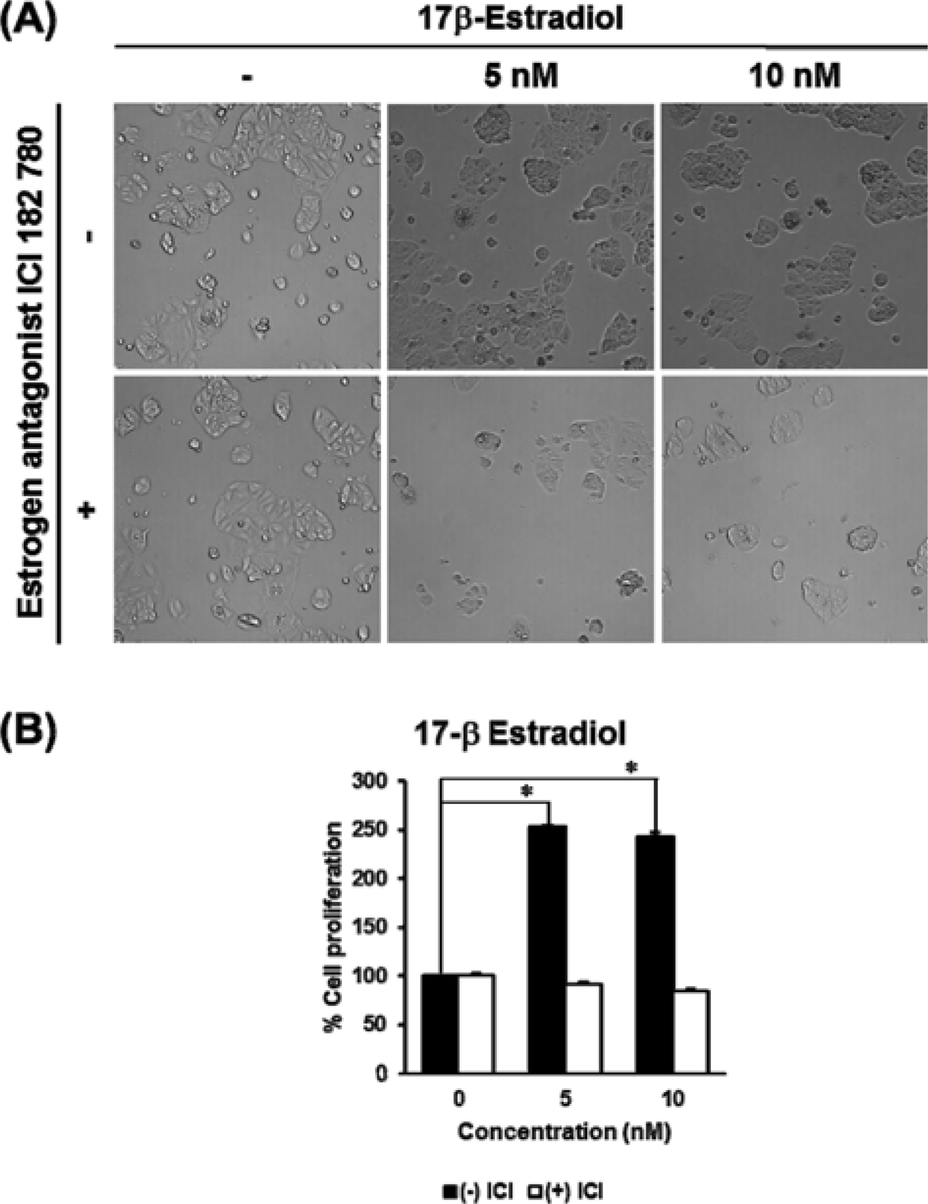
Fig. 1. The estrogenic effect of 17β-estradiol on the proliferation of MCF-7 human breast cancer cells. (A) The microscopic pictures from E-screen assay results of 17β-estradiol. (B) The comparative graph illustrates the percentage increase in cell proliferation compared with the untreated group. In this E-screen assay, 17β-estradiol was used as a positive control. MCF-7 cells were incubated in 24-well plates and treated with tested sample in phenol red-free RPMI medium supplemented with 5% charcoal-dextran-stripped human for 144 hours. For the antagonistic test, the pure estrogen receptor antagonist ICI 182 780 was added with the test materials. Ez-Cytox reagent was added to each well for 1hour and the cell viability was then calculated from the measure-ment of the optical density at 450 nm using a microplate reader. P values of less than 0.001 were considered statistically significant.
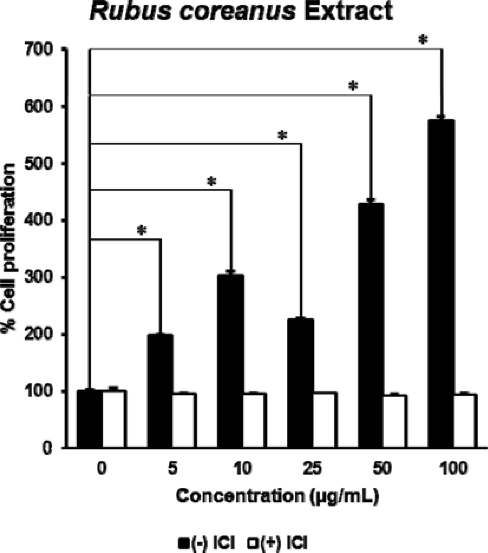
Fig. 2. The estrogenic effect of Rubus coreanus extract on the proliferation of MCF-7 cells. The extract of R. coreanus significantly stimulated cell growth in a dose-dependent manner, especially at the concentration of 100 μ g/mL. P values of less than 0.001 were considered statistically significant.
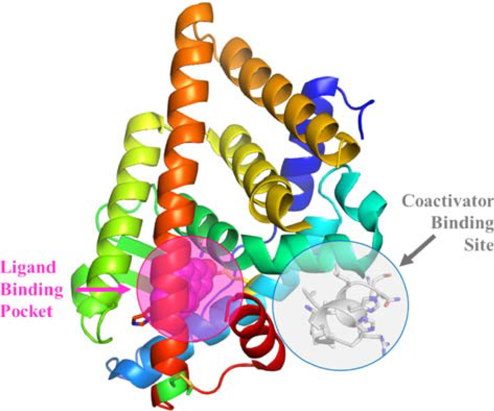
Fig. 3. Structure of ligand-binding domain of estrogen receptor α with the ligand-binding pocket and coactivator-binding site. Not only the binding between estrogen and its ligand binding pocket, the protein-protein interactions between coactivator proteins and their binding sites also results in cell proliferation. Competitive blockade of the coactivator binding site could effectively halt cell proliferation for breast cancer therapeutics.
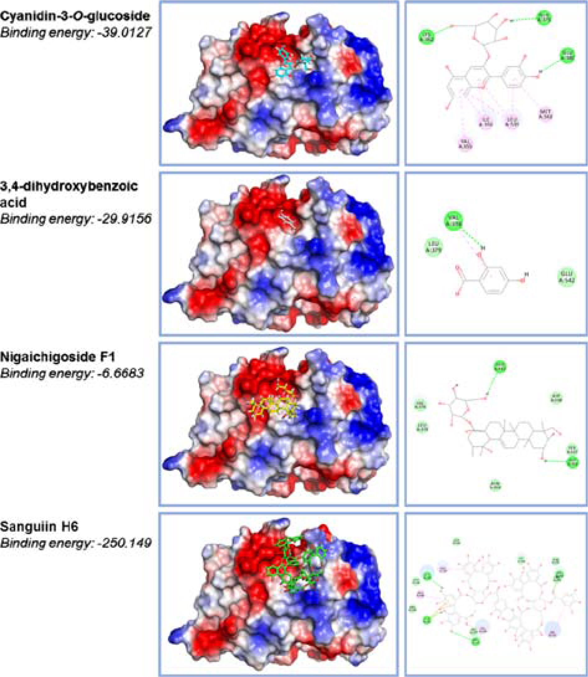
Fig. 4. The docking structures of R. coreanus compounds with coactivator-binding site of estrogen receptor α. Molecular docking assay was constructed from the X-ray structure of ERα complexed with estradiol and a synthesized stable peptide inhibitor in the coactivator-binding groove. The atomic coordinates were obtained from the Protein Data Bank and manipulated using Discovery Studio molecular modeling package. The docking simulation was implemented by CDOCKER program. All atomic charges of ligands were assigned using the Momany-Rone partial charge and CHARMM force fields. Among four compounds isolated from R. coreanus, Sanguiin H6 showed the strongest affinity for the ERα coactivator-binding site.
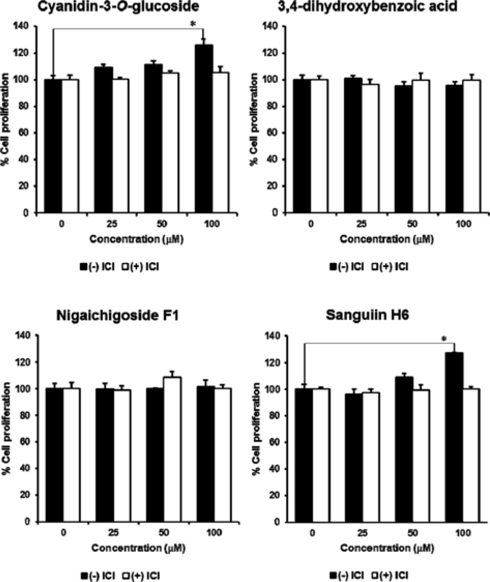
Fig. 5. The estrogenic effects of R. coreanus compounds on the proliferation of MCF-7 cells. Sanguiin H6 and cyanidin-3-O-glucoside slightly induced the proliferation of MCF-7 cells at the concentration of 100 μM. P values of less than 0.001 were considered statistically significant.
Reference
-
(1). Oh C. M., Won Y. J., Jung K. W., Kong H. J., Cho H., Lee J. K., Lee D. H., Lee K. H.Cancer Res. Treat. 2016; 48:436–450.(2). Jung K. W., Won Y. J., Oh C. M., Kong H. J., Lee D. H., Lee K. H.Cancer Res. Treat. 2017; 49:306–312.(3). Carmichael A. R., Mokbel K.Arch. Plast. Surg. 2016; 43:222–223.(4). Irelli A., Cocciolone V., Cannita K., Zugaro L., Di Staso M., Lanfiuti Baldi P. L., Paradisi S., Sidoni T., Ricevuto E., Ficorella C.Bone. 2016; 87:169–175.(5). de Pedro M., Baeza S., Escudero M. T., Dierssen-Sotos T., Gómez-Acebo I., Pollán M., Llorca J.Breast Cancer Res. Treat. 2015; 149:525–536.(6). Esteva F. J., Hortobagyi G. N.Sci. Am. 2008; 298:58–65.(7). Jameera Begam A., Jubie S., Nanjan M. J.Bioorg. Chem. 2017; 71:257–274.(8). Howell S. J., Johnston S. R. D., Howell A.Best Pract. Res. Clin. Endocrinol. Metab. 2004; 18:47–66.(9). Zheng J., Zhou Y., Li Y., Xu D. P., Li S., Li H. B.Nutrients. 2016; 8:495.(10). Lee J., Dossett M., Finn C. E.Molecules. 2014; 19:10524–10533.(11). Heo J.Donguibogam; Yeogang: Korea. 1994; 946–947.(12). Li J., Du L. F., He Y., Yang L., Li Y. Y., Wang Y. F., Chai X., Zhu Y., Gao X. M.Chem. Biodivers. 2015; 12:1809–1847.(13). Ju H. K., Cho E. J., Jang M. H., Lee Y. Y., Hong S. S., Park J. H., Kwon S. W. J.Pharm. Biomed. Anal. 2009; 49:820–827.(14). Choung M. G., Lim J. D.Korean J. Med. Crop Sci. 2012; 20:259–269.269.(15) Körner W.., Hanf V.., Schuller W.., Kempter C.., Metzqer J.., Haqenmaier H.Sci. Total Environ. 1999. 225:33–48.(16). Soto A. M., Sonnenschein C., Chung K. L., Fernandez M. F., Olea N., Serrano F. O.Environ. Health Perspect. 1995; 103:113–122.(17). Lee S., Barron M. G.PloS One. 2017; 12:1–14.(18). Ng H. W., Zhang W., Shu M., Luo H., Ge W., Perkins R., Tong W., Hong H.BMC Bioinformatics. 2014; 15:1–15.(19). Pang X., Fu W., Wang J., Kang D., Xu L., Zhao Y., Liu A. L., Du G. H.Oxid. Med. Cell. Longev. 2018; 2018:1–11.(20). Jordan V. C. J.Med. Chem. 2003; 46:883–908.(21). McDonnell D. P., Chang C. Y., Norris J. D. J.Steroid Biochem. Mol. Biol. 2000; 74:327–335.(22). Sun A., Moore T. W., Gunther J. R., Kim M. S., Rhoden E., Du Y., Fu H., Snyder J. P., Katzenellenbogen J. A.Chem. Med. Chem. 2011; 6:654–666.(23). Park E. J., Lee D., Baek S. E., Kim K. H., Kang K. S., Jang T. S., Lee H. L., Song J. H., Yoo J. E.Bioorg. Med. Chem. Lett. 2017; 27:4389–4392.(24). Park E. H., Park J. Y., Yoo H. S., Yoo J. E., Lee H. L.Bioorg. Med. Chem. Lett. 2016; 26:3291–3294.(25). Choi M. H., Shim S. M., Kim G. H. J.Food Sci. Technol. 2016; 53:1214–1221.(26). Ko H., Jeon H., Lee D., Choi H. K., Kang K. S., Choi K. C.Bioorg. Med. Chem. Lett. 2015; 25:5508–5513.(27). Helferich W. G., Andrade J. E., Hoagland M. S.Inflammophar-macology. 2008; 16:219–226.(28). Hsieh C. Y., Santell R. C., Haslam S. Z., Helferich W. G.Cancer Res. 1998; 58:3833–3838.(29). Lee J. Y., Kim H. S., Song Y. S. J.Tradit. Complement. Med. 2012; 2:96–104.
- Full Text Links
-
- Actions
-
Cited
- CITED
-
- Close
- Share
-
- Similar articles
-
- Additive Estrogenic Activities of the Binary Mixtures of Four Estrogenic Chemicals in Recombinant Yeast Expressing Human Estrogen Receptor
- Selective Estrogen Receptor Modulators: A Review of Action Mechanism and Clinical Data
- Requirement of Metabolic Activation for Estrogenic Activity of Pueraria mirifica
- Sterol-independent repression of low density lipoprotein receptor promoter by peroxisome proliferator activated receptor gamma coactivator-1alpha (PGC-1alpha)
- Effects of estrogen receptor and estrogen on the chromatin structure in estrogen receptor stable transfectants
