Anti-inflammatory Activity of Standardized Fraction from Inula helenium L. via Suppression of NF-κB Pathway in RAW 264.7 Cells
- Affiliations
-
- 1Natural Products Research Institute, College of Pharmacy, Seoul National University, Seoul 08826, Republic of Korea. kims@snu.ac.kr
- 2Current Address: Winship Cancer Institute, Emory University School of Medicine, Atlanta, GA 30322, USA.
- KMID: 2443098
- DOI: http://doi.org/10.20307/nps.2019.25.1.16
Abstract
- Inula helenium L. is rich source of eudesmane-type sesquiterpene lactones, mainly alantolactone and isoalantolactone, which have the various pharmacological functions. In this study, we examined the inhibitory effects of nitric oxide (NO) production of hexane, methylene chloride, ethyl acetate, butanol, and water fractions from I. helenium and investigated the anti-inflammatory effect of hexane fraction of I. helenium (HFIH) in LPS-induced RAW 264.7 cells. Quantification of alantolactone and isoalantolactone from HFIH was carried out for the standardization by multiple reaction monitoring using triple quadrupole mass spectrometer. HFIH significantly inhibited inducible NO synthase (iNOS) and cyclooxygenase-2 (COX-2) protein as well as their downstream products NO and prostaglandin E₂ (PGE₂) in LPS-stimulated RAW 264.7 cells. Moreover, HFIH suppressed NF-κB transcriptional activity by decreasing the translocation of p65 to the nucleus. The in vivo study further confirmed that HFIH attenuated the paw edema induced by carrageenan in an acute inflammation model. These findings suggest that HFIH may be useful as a promising phytomedicine for inflammatory-associated diseases.
MeSH Terms
Figure
-
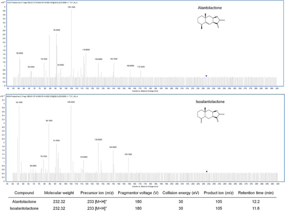
Fig. 1 Chemical structures of alantolactone and isoalantolactone and product ion scan for multiple reaction monitoring (MRM). The selected m/z transition for alantolactone and isoalantolactone was 233 →105. MRM parameters were listed.
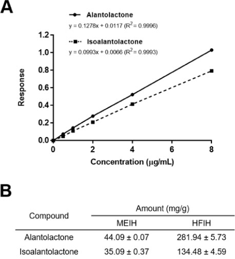
Fig. 2 Quantification of alantolactone and isoalantolactone by MRM mode of triple quadrupole mass spectrometry. (A) Calibration curves of alantolactone and isoalantolactone by MRM analysis. (B) Calculated amount of alantolactone and isoalantolactone in MEIH and HFIH.
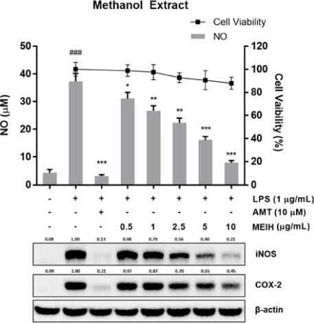
Fig. 3 Inhibitory effect of methanol extract of I. helenium (MEIH) on NO production and iNOS and COX-2 expression in LPS-induced RAW 264.7 cells. Cells were pretreated with MEIH for 2 h and stimulated for 18 h with LPS. Anti-proliferative effect was determined using MTT assay. The amount of NO in the medium was measured using a Griess reagent. The expression of iNOS and COX-2 was determined by Western blotting. The results are represented as the mean ± SD (n = 3) from three independent experiments. ###p < 0.001 indicates significant difference from the unstimulated control group. *p < 0.05, **p < 0.01, and ***p < 0.001 indicated the significant difference compared to LPS-treated control group.
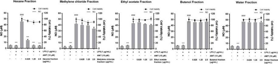
Fig. 4 Inhibitory effects of fractions from MEIH on NO production in LPS-induced RAW 264.7 cells. Cells were treated with various concentrations of fractions (0 – 2.5 µg/mL). Effects of fractions on NO production were measured by Griess reagent. The results are represented as the mean ± SD (n = 3) from three independent experiments. ###p < 0.001 indicates significant difference from the unstimulated control group. ***p < 0.001 indicated the significant difference compared to LPS-treated control group.

Fig. 5 Inhibitory effect of the hexane fraction of I. helenium (HFIH) on the production of NO and PGE2 and the expression of iNOS and COX-2 in LPS-induced RAW 264.7 cells. Cells were pretreated with HFIH for 2 h and stimulated for 18 h with LPS. The amounts of NO and PGE2 in the medium were measured using a Griess reagent and PGE2 ELISA kit. The expression of iNOS and COX-2 was determined by Western blotting. The results are represented as the mean ± SD (n = 3) from three independent experiments. ###p < 0.001 indicates significant difference from the unstimulated control group. *p < 0.05, **p < 0.01, and ***p < 0.001 indicated the significant difference compared to LPS-treated control group.
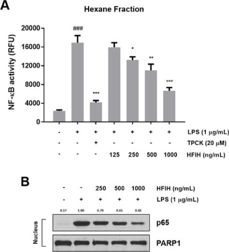
Fig. 6 Inhibitory effect of NF-κB activation in LPS-stimulated RAW 264.7 cells. (A) Cells harboring the NF-κB SEAP-NPT reporter construct were pretreated with the indicated concentrations of HFIH for 2 h then exposed to LPS. The SEAP activity was measured in RFU, using a microplate fluorometer. (B) Effect of HFIH on NF-κB p65 localization to the nucleus. Nuclear cell lysates were prepared for Western blotting. The results are represented as the mean ± SD (n = 3) from three independent experiments. ###p < 0.001 indicates significant difference from the unstimulated control group. *p < 0.05, **p < 0.01, and ***p < 0.001 indicated the significant difference compared to LPS-treated control group.
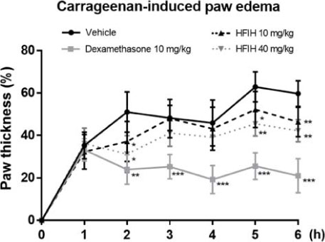
Fig. 7 Inhibitory effect of HFIH on carrageenan-induced paw edema mice model. HFIH (10 and 40 mg/kg) was injected intraperitoneally 2 h prior to 1% carrageenan administration into the right hind paw. 6 mice per each group were used in this study. Paw thickness was measured every hour for 6 h with a dial thickness gauge. Percent increase in paw thickness was calculated by measuring the difference between the control paw and the carrageenan-induced paw at each point. Dexamethasone (10 mg/kg) was used as a positive control. The results are represented as the mean ± SD (n = 6). *p < 0.05, **p < 0.01, and ***p < 0.001 indicated the significant difference compared to carrageenan-injected control group.

Fig. 8 Inhibitory effects of alantolactone (A) and isoalantolactone (B) on the production of NO and PGE2 and the expression of iNOS and COX-2 in LPS-induced RAW 264.7 cells. Cells were pretreated with compounds for 2 h and stimulated for 18 h with LPS. The amounts of NO and PGE2 in the medium were measured using a Griess reagent and PGE2 ELISA kit. The expression of iNOS and COX-2 was determined by Western blotting. The results are represented as the mean ± SD (n = 3) from three independent experiments. ###p < 0.001 indicates significant difference from the unstimulated control group. ***p < 0.001 indicated the significant difference compared to LPS-treated control group.
Reference
-
1. Fujiwara N, Kobayashi K. Curr Drug Targets Inflamm Allergy. 2005; 4:281–286.2. Amin AR, Attur M, Abramson SB. Curr Opin Rheumatol. 1999; 11:202–209.3. Tak PP, Firestein GS. J Clin Invest. 2001; 107:7–11.4. Yamamoto Y, Gaynor RB. Trends Biochem Sci. 2004; 29:72–79.5. Yuan G, Wahlqvist ML, He G, Yang M, Li D. Asia Pac J Clin Nutr. 2006; 15:143–152.6. Spiridon I, Nechita CB, Niculaua M, Silion M, Armatu A, Teaca CA, Bodirlau R. Cent Eur J Chem. 2013; 11:1699–1709.7. Trendafilova A, Chanev C, Todorova M. Pharmacogn Mag. 2010; 6:234–237.8. Babaei G, Aliarab A, Abroon S, Rasmi Y, Aziz SG. Biomed Pharmacother. 2018; 106:239–246.9. Chun J, Choi RJ, Khan S, Lee DS, Kim YC, Nam YJ, Lee DU, Kim YS. Int Immunopharmacol. 2012; 14:375–383.10. He G, Zhang X, Chen Y, Chen J, Li L, Xie Y. Biomed Pharmacother. 2017; 90:598–607.11. Ketai W, Huitao L, Yunkun Z, Xingguo C, Zhide H, Yucheng S, Xiao M. Talanta. 2000; 52:1001–1005.12. Gao S, Wang Q, Tian XH, Li HL, Shen YH, Xu XK, Wu GZ, Hu ZL, Zhang WD. J Ethnopharmacol. 2017; 196:39–46.13. Engström MT, Pälijärvi M, Salminen JP. J Agric Food Chem. 2015; 63:4068–4079.14. Ahn KS, Noh EJ, Zhao HL, Jung SH, Kang SS, Kim YS. Life Sci. 2005; 76:2315–2328.15. Kim SF, Huri DA, Snyder SH. Science. 2005; 310:1966–1970.16. Tang X, Liu D, Shishodia S, Ozburn N, Behrens C, Lee JJ, Hong WK, Aggarwal BB, Wistuba II. Cancer. 2006; 107:2637–2646.17. Morris CJ. Methods Mol Biol. 2003; 225:115–121.18. Guo C, Zhang S, Teng S, Niu K. J Sep Sci. 2014; 37:950–956.19. Calixto JB. Braz J Med Biol Res. 2000; 33:179–189.
- Full Text Links
-
- Actions
-
Cited
- CITED
-
- Close
- Share
-
- Similar articles
-
- Anti-inflammatory effects of proanthocyanidin-rich red rice extract via suppression of MAPK, AP-1 and NF-κB pathways in Raw 264.7 macrophages
- Luteolin 5-O-glucoside from Korean Milk Thistle, Cirsium maackii, Exhibits Anti-Inflammatory Activity via Activation of the Nrf2/HO-1 Pathway
- Anti-inflammatory activities of Scolopendra subspinipes mutilans in RAW 264.7 cells
- Rhodanthpyrone A and B play an anti-inflammatory role by suppressing the nuclear factor-κB pathway in macrophages
- Estragole Exhibits Anti-inflammatory Activity with the Regulation of NF-κB and Nrf-2 Signaling Pathways in LPS-induced RAW 264.7 cells
