Diffusion Tensor Imaging and Quantitative Magnetic Resonance Volumetric Assessment in the Diagnosis of Hemimegalencephaly: A Case Report
- Affiliations
-
- 1Department of Radiology, Hanyang University Hospital, Hanyang University College of Medicine, Seoul, Korea. jyjy133@naver.com
- 2Department of Radiology, Hanyang University Guri Hospital, Hanyang University College of Medicine, Guri, Korea.
- 3Department of Neurology, Hanyang University Hospital, Hanyang University College of Medicine, Seoul, Korea.
- KMID: 2442478
- DOI: http://doi.org/10.3348/jksr.2019.80.1.164
Abstract
- A 27-year-old female presented with repeated seizures. As the left frontal lobe volume was enlarged in comparison with the right frontal lobe, hemimegalencephaly was suggested. Abnormal white matter fiber tracts running from the left frontal lobe to the fornix and hippocampus were found on diffusion tensor imaging (DTI). We performed quantitative measurements of brain volume and confirmed hemimegalencephaly. DTI and MRI-based volumetry techniques could be useful to objectively diagnose hemimegalencephaly.
MeSH Terms
Figure
-
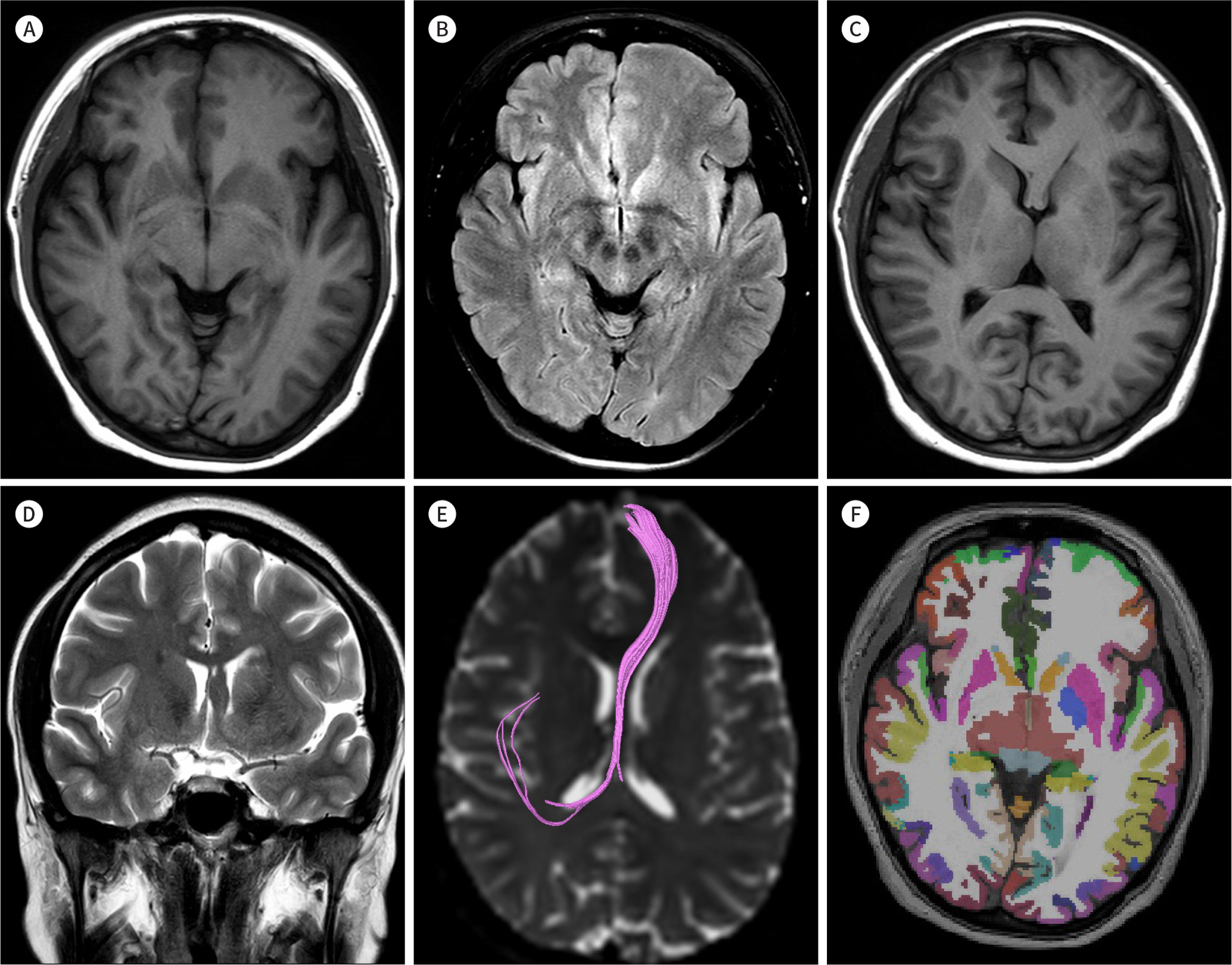
Fig. 1. A 27-year-old female with feature of hemimegalencephaly on conventional MRI (A-D), DTI (E), and MR volumetry (F). Axial T1-weighted (A) and T2 fluid-attenuated inversion recovery (B) images show asymmetrically broadened gyri of the left frontal lobe with blurring of gray-white matter differentiation. Axial T1-weighted (C) and coronal T2-weighted (D) images demonstrate asymmetric thickened left fornix, which is iso-signal intensity to white matter and mildly compressing the frontal horn of the left lateral ventricle. Aberrant and asymmetric white matter fiber tracts running from the left frontal lobe to the fornix and hippocampus are observed on DTI (E). For MR volumetry, Destrieux atlas based automatic parcellation of cortical surface is overlaied on T1-weighted image (F). DTI = diffusion tensor imaging
Reference
-
References
1. Flores-Sarnat L. Hemimegalencephaly: part 1. Genetic, clinical, and imaging aspects. J Child Neurol. 2002; 17:373–384. ; discussion 384.
Article2. Sato N, Ota M, Yagishita A, Miki Y, Takahashi T, Adachi Y, et al. Aberrant midsagittal fiber tracts in patients with hemimegalencephaly.AJNR Am J Neuroradiol. 2008; 29:823–827.3. Takahashi T, Sato N, Ota M, Nakata Y, Yamashita F, Adachi Y, et al. Asymmetrical interhemispheric fiber tracts in patients with hemimegalencephaly on diffusion tensor magnetic resonance imaging.J Neuroradiol. 2009; 36:249–254.4. Farid N, Girard HM, Kemmotsu N, Smith ME, Magda SW, Lim WY, et al. Temporal lobe epilepsy: quantitative MR volumetry in detection of hippocampal atrophy. Rad/iiology. 2012; 264:542–550.
Article5. Huang H, Zhang J, Jiang H, Wakana S, Poetscher L, Miller MI, et al. DTI tractography based parcellation of white matter: application to the midsagittal morphology of corpus callosum.Neuroimage. 2005; 26:195–205.6. Desikan RS, Ségonne F, Fischl B, Quinn BT, Dickerson BC, Blacker D, et al. An automated labeling system for subdividing the human cerebral cortex on MRI scans into gyral based regions of interest.Neuroimage. 2006; 31:968–980.7. Destrieux C, Fischl B, Dale A, Halgren E. Automatic parcellation of human cortical gyri and sulci using standard anatomical nomenclature.Neuroimage. 2010; 53:1–15.8. Santos AC, Escorsi-Rosset S, Simao GN, Terra VC, Velasco T, Neder L, et al. Hemispheric dysplasia and hemimegalencephaly: imaging definitions. Childs Nerv Syst. 2014; 30:1813–1821.
Article9. Atul A, Gautam D, Daljit S, Deepashu S, Robin G. Hemimegalencephaly without seizures: report of a case and review of literature. Romanian Neurosurg. 2017; 31:306–309.
Article10. Beaulieu-Boire I, Lortie A, Bissonnette J, Prevost S, Bergeron D, Bocti C. Hemimegalencephaly in an adult with normal intellectual function and mild epilepsy. Dev Med Child Neurol. 2012; 54:284–286.
Article
- Full Text Links
-
- Actions
-
Cited
- CITED
-
- Close
- Share
-
- Similar articles
-
- Principle and Experiments in Diffusion Tensor Imaging
- Structural MR Imaging in the Diagnosis of Alzheimer's Disease and Other Neurodegenerative Dementia: Current Imaging Approach and Future Perspectives
- Diffusion Tensor Imaging: Exploring the Motor Networks and Clinical Applications
- Diffusion Tensor Tractography of a Gliomatosis Cerebri: A Case Report
- Delayed Anoxic Encephalopathy after Carbon Monoxide Poisoning: Evaluation of Therapeutic Effect by Serial Diffusion-Tensor Magnetic Resonance Imaging and Neurocognitive Test
