J Korean Soc Radiol.
2019 Jan;80(1):105-116. 10.3348/jksr.2019.80.1.105.
Dual-Layer Spectral Detector CT Discography of the Lumbar Spine: A Preliminary Study
- Affiliations
-
- 1Department of Radiology, Seoul National University Hospital, Seoul, Korea. drhong@snu.ac.kr
- 2Department of Radiology, National Health Insurance Service Ilsan Hospital, Goyang, Korea.
- KMID: 2442469
- DOI: http://doi.org/10.3348/jksr.2019.80.1.105
Abstract
- PURPOSE
To assess the feasibility of spectral detector CT (SDCT) with axial maximum-intensity projection (MIP) reconstruction for the evaluation of lumbar CT discography.
MATERIALS AND METHODS
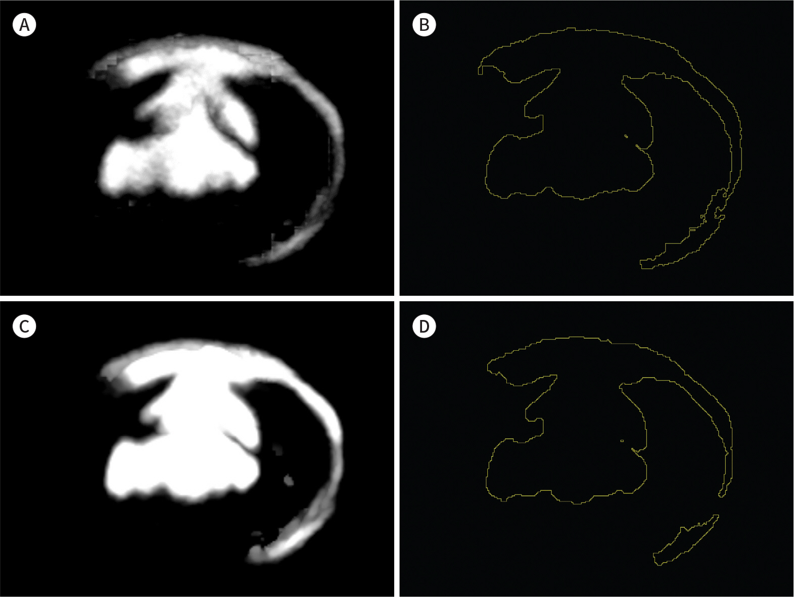
We retrospectively evaluated 44 disc levels from 18 patients who underwent CT discography on a dual-layer SDCT between May 2016 and July 2017. We compared the distribution of contrast material between conventional CT and SDCT-based iodine maps using the Jaccard index (JI) and Dice similarity coefficient (DSC). Qualitative analysis of the post-discogram features was done according to the Dallas discogram description, and changes in reading time and diagnostic confidence were analyzed.
RESULTS
The intermethod variability between conventional CT and SDCT was good, with a mean DSC of 0.93 and a mean JI of 0.87. The mean sensitivity and positive predictive value of the SDCT-based method were 90% and 96%, respectively. The addition of SDCT-based axial MIP iodine maps increased the diagnostic confidence (p = 0.025) and reduced the reading time in both reviewers (p < 0.001).
CONCLUSION
SDCT discography demonstrates the distribution of contrast medium within the disc similarly to conventional CT. Additionally, axial MIP iodine maps using SDCT allow for the fast evaluation of disc pathology with reduced reading time and can increase diagnostic confidence.
MeSH Terms
Figure
Reference
-
References
1. Saboeiro GR. Lumbar discography. Radiol Clin North Am. 2009; 47:421–433.2. Manchikanti L, Benyamin RM, Singh V, Falco FJ, Hameed H, Derby R, et al. An update of the systematic appraisal of the accuracy and utility of lumbar discography in chronic low back pain. Pain Physician. 2013; 16:SE55–SE95.3. Myung JS, Lee JW, Park GW, Yeom JS, Choi JY, Hong SH, et al. MR diskography and CT diskography with gadodiamide-iodinated contrast mixture for the diagnosis of foraminal impingement. AJR Am J Roentgenol. 2008; 191:710–715.4. Kaza RK, Ananthakrishnan L, Kambadakone A, Platt JF. Update of dualenergy CT applications in the genitourinary tract. AJR Am J Roentgenol. 2017; 208:1185–1192.5. Johnson TR. Dual-energy CT: general principles. AJR Am J Roentgenol. 2012; 199:S3–S8.6. Bongartz T, Glazebrook KN, Kavros SJ, Murthy NS, Merry SP, Franz WB 3rd, et al. Dual-energy CT for the diagnosis of gout: an accuracy and diagnostic yield study. Ann Rheum Dis. 2015; 74:1072–1077.7. Kaup M, Wichmann JL, Scholtz JE, Beeres M, Kromen W, Albrecht MH, et al. Dual-energy CT-based display of bone marrow edema in osteoporotic vertebral compression fractures: impact on diagnostic accuracy of radiologists with varying levels of experience in correlation to MR imaging.Radiology. 2016; 280:510–519.8. Glazebrook KN, Brewerton LJ, Leng S, Carter RE, Rhee PC, Murthy NS, et al. Case-control study to estimate the performance of dualenergy computed tomography for anterior cruciate ligament tears in patients with history of knee trauma.Skeletal Radiol. 2014; 43:297–305.9. Dice LR. Measures of the amount of ecologic association between species.Ecology. 1945; 26:297–302.10. Bakic PR, Carton AK, Kontos D, Zhang C, Troxel AB, Maidment AD. Breast percent density: estimation on digital mammograms and central tomosynthesis projections. Radiology. 2009; 252:40–49.
Article11. Thévenaz P, Ruttimann UE, Unser M. A pyramid approach to subpixel registration based on intensity.IEEE Trans Image Process. 1998; 7:27–41.12. Heras J. DetectionEvaluationJ Available at. https://joheras.github.io/DetectionEvaluationJ. Accessed Jun 27,. 2018.13. Sachs BL, Vanharanta H, Spivey MA, Guyer RD, Videman T, Rashbaum RF, et al. Dallas discogram description. A new classification of CT/discography in low-back disorders. Spine (Phila Pa 1976). 1987; 12:287–294.
Article14. Bagci AM, Lee SH, Nagornaya N, Green BA, Alperin N. Automated posterior cranial fossa volumetry by MRI: applications to Chiari malformation type I. AJNR Am J Neuroradiol. 2013; 34:1758–1763.
Article15. Fleiss JL, Levin B, Cho Paik M.Statistical methods for rates and proportions. 3rd ed.New York, NY: John Wiley & Sons;2013.16. Chou R, Loeser JD, Owens DK, Rosenquist RW, Atlas SJ, Baisden J, et al. Interventional therapies, surgery, and interdisciplinary rehabilitation for low back pain: an evidencebased clinical practice guideline from the American Pain Society.Spine (Phila Pa 1976). 2009; 34:1066–1077.17. Cuellar JM, Stauff MP, Herzog RJ, Carrino JA, Baker GA, Carragee EJ. Does provocative discography cause clinically important injury to the lumbar intervertebral disc? A 10-year matched cohort study.Spine J. 2016; 16:273–280.18. Bernard TN Jr. Lumbar discography followed by computed tomography. Refining the diagnosis of low-back pain.Spine (Phila Pa 1976). 1990; 15:690–707.
Article19. Manchikanti L, Glaser SE, Wolfer L, Derby R, Cohen SP. Systematic review of lumbar discography as a diagnostic test for chronic low back pain. Pain Physician. 2009; 12:541–559.20. Schellhas KP, Pollei SR, Gundry CR, Heithoff KB. Lumbar disc high-intensity zone. Correlation of magnetic resonance imaging and discography.Spine (Phila Pa 1976). 1996; 21:79–86.
Article21. Johnson T, Fink C, Schönberg SO, Reiser MF.Dual energy CT in clinical practice. Berlin: Springer;2011.22. Patino M, Prochowski A, Agrawal MD, Simeone FJ, Gupta R, Hahn PF, et al. Material separation using dualenergy CT: current and emerging applications.Radiographics. 2016; 36:1087–1105.23. Jun WS. Dual-energy CT discography: experimental study for validation of automated bone removal application [dissertation]. Reston: Seoul National University;2009.24. Mallinson PI, Coupal T, Reisinger C, Chou H, Munk PL, Nicolaou S, et al. Artifacts in dualenergy CT gout protocol: a review of 50 suspected cases with an artifact identification guide. AJR Am J Roentgenol. 2014; 203:W103–W109.
- Full Text Links
- Actions
-
Cited
- CITED
-
- Close
- Share
- Similar articles
-
- Spectral CT Multiparameter Angiography in the Diagnosis of Aortic Stent Endoleak
- CT Scan and Discographic Findings in Ruptured Lumbar Discs
- Significance of CT after discography
- CT-Discography: Diagnostic Accuracy in Lumbar Disc Herniation and Significance of Induced Pain During Procedure
- Utility of Discography as a Preoperative Diagnostic Tool for Intradural Lumbar Disc Herniation


