Anat Cell Biol.
2019 Mar;52(1):102-104. 10.5115/acb.2019.52.1.102.
An elongated dorsally curved xiphoid process
- Affiliations
-
- 1Seattle Science Foundation, Seattle, WA, USA. joei@seattlesciencefoundation.org
- 2Division of Gross and Clinical Anatomy, Department of Anatomy, Kurume University School of Medicine, Kurume, Fukuoka, Japan.
- 3Department of Anatomical Sciences, St. George's University, St. George's, Grenada, West Indies.
- KMID: 2442340
- DOI: http://doi.org/10.5115/acb.2019.52.1.102
Abstract
- The xiphoid process of the sternum lies in the epigastric region and functions to serve as an attachment point for vital muscles that aid in respiration. With the xiphoid process extending as the most inferior portion of the sternum, variable morphology is widely observed. During a routine dissection of a 44-year-old Caucasian male cadaver, we discovered a hook-shaped, elongated xiphoid process that protruded dorsally. Potential clinical significance can arise leading to misdiagnosis of the hook-shaped xiphoid process as an epigastric mass during imaging. Though various variations of xiphoid process have been well documented, knowledge of a hook-shaped xiphoid process orientated dorsally remains scarce. Herein, this case study provides clinicians, surgeons, and radiologists a rare anomaly of the xiphoid process in order to further the knowledge of morphological variations of the xiphoid to prevent misdiagnosis and surgical complications.
Keyword
MeSH Terms
Figure
-
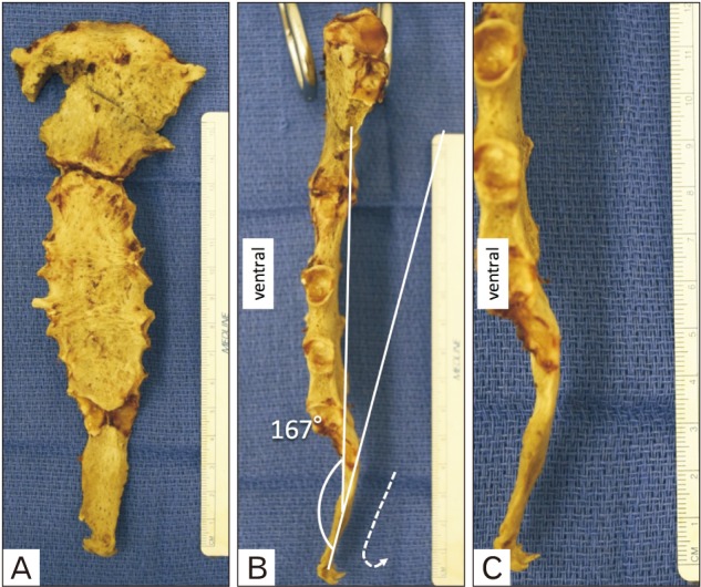
Fig. 1 Variant of the xiphoid process. (A) An anterior view of the sternum. (B) Lateral view illustrating the angle between the sternal body and xiphoid process. Note the hook-shaped xiphoid process. (C) Lateral view with a scale.
Reference
-
1. Voin V, Oskouian RJ, Loukas M, Tubbs RS. Auscultation of the heart: the basics with anatomical correlation. Clin Anat. 2017; 30:58–60. PMID: 27576554.2. Graeber GM, Nazim M. The anatomy of the ribs and the sternum and their relationship to chest wall structure and function. Thorac Surg Clin. 2007; 17:473–489. PMID: 18271162.3. Nagasao T, Kasai S, Shimizu Y, Sakamoto Y, Hatano A, Morotomi T, Ogata H, Kishi K. A biomechanical study of relationship between sternum defect patterns and thoracic respiration. Comput Assist Surg (Abingdon). 2016; 21:1–8. PMID: 27973954.4. Simpson JK, Hawken E. Xiphodynia: a diagnostic conundrum. Chiropr Osteopat. 2007; 15:13. PMID: 17868466.5. Xie YZ, Wang BJ, Yun JS, Chung GH, Ma ZB, Li XJ, Kim IS, Chai OH, Han EH, Kim HT, Song CH. Morphology of the human xiphoid process: dissection and radiography of cadavers and MDCT of patients. Surg Radiol Anat. 2014; 36:209–217. PMID: 23839070.6. Standring S. Gray's anatomy: the anatomical basis of clinical practice. 41st ed. New York: Elsevier;2016. p. 932–934.7. Eid S, Iwanaga J, Oskouian RJ, Loukas M, Tubbs RS. Bifid xiphoid process: case report and review. Cureus. 2018; 10:e3153. PMID: 30345208.8. Yekeler E, Tunaci M, Tunaci A, Dursun M, Acunas G. Frequency of sternal variations and anomalies evaluated by MDCT. AJR Am J Roentgenol. 2006; 186:956–960. PMID: 16554563.9. Akin K, Kosehan D, Topcu A, Koktener A. Anatomic evaluation of the xiphoid process with 64-row multidetector computed tomography. Skeletal Radiol. 2011; 40:447–452. PMID: 20721551.10. Ateşoğlu S, Deniz M, Uslu AI. Evaluation of the morphological characteristic and sex differences of sternum by multi-detector computed tomography. Folia Morphol (Warsz). 2018; 77:489–497. PMID: 29345718.11. Mashriqi F, D'Antoni AV, Tubbs RS. Xiphoid process variations: a review with an extremely unusual case report. Cureus. 2017; 9:e1613. PMID: 29098125.12. Fischer NJ, Morreau J, Sugunesegran R, Taghavi K, Mirjalili SA. A reappraisal of pediatric thoracic surface anatomy. Clin Anat. 2017; 30:788–794. PMID: 28514496.13. Pak N, Patel SG, Hashemi Taheri AP, Hashemi F, Eftekhari Vaghefi R, Naybandi Atashi S, Mirjalili SA. A reappraisal of adult thoracic and abdominal surface anatomy in Iranians in vivo using computed tomography. Clin Anat. 2016; 29:191–196. PMID: 26575429.14. Keough N, Mirjalili SA, Suleman FE, Lockhat ZI, van Schoor A. The thoracic surface anatomy of adult black South Africans: a reappraisal from CT scans. Clin Anat. 2016; 29:1018–1024. PMID: 27571396.
- Full Text Links
-
- Actions
-
Cited
- CITED
-
- Close
- Share
-
- Similar articles
-
- Heterotopic Ossification of the Xiphoid Process after Abdominal Surgery for Traumatic Hemoperitoneum
- A Xiphoid Elongation Following a Trauma Laparotomy: A Case Report
- Xiphodynia: 2 cases report
- A rare triple skeletal bone variation, including a median cleft of the posterior arch of the atlas, a sternal foramina and a bifid xiphoid process
- Three Dimensional Images of the Sternum in Children with Using MDCT
