Anat Cell Biol.
2019 Mar;52(1):87-89. 10.5115/acb.2019.52.1.87.
Superficial course of the medial plantar nerve: case report
- Affiliations
-
- 1Department of Anatomy, Yeungnam University College of Medicine, Daegu, Korea. jookim@med.yu.ac.kr
- 2Department of Anatomy, Keimyung University School of Medicine, Daegu, Korea.
- KMID: 2442335
- DOI: http://doi.org/10.5115/acb.2019.52.1.87
Abstract
- The medial and lateral plantar nerves are branched from the tibial nerve and move to the tip of the toes. A variation of medial plantar nerve was found on the left side of a 78-year-old Korean male cadaver. The tibial nerve was divided into the lateral and medial plantar nerves beneath the plantar flexor. The medial plantar nerve passed deep to plantar aponeurosis and superficial to the flexor digitorum brevis. It gave off a common plantar digital nerve and then divided into three proper plantar digital nerves near the metatarsal bases. In this article, we report a superficial course of the medial plantar nerve and describe its unique morphology and discuss the clinical significance of this variation.
Keyword
Figure
-
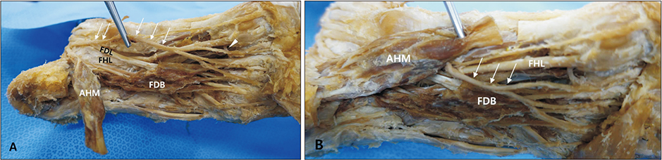
Fig. 1 Photograph of variant course of the medial plantar nerve. (A) After cutting abductor hallucis muscle (AHM), the medial plantar nerve (arrows) continued superficial to flexor digitorum brevis (FDB). And its branches accompanied the branches of the medial plantar artery (arrowhead). (B) Superficial course of the branches of the medial plantar artery (arrows). FDL, flexor digitorum longus; FHL, flexor hallucis longus.
Cited by 1 articles
-
Anatomical variation of quadratus plantae in relation with flexor digitorum brevis
Punnapa Raviteja, Mrudula Chandrupatla, Ramoju Harshitha, Marni Sameer Chowdary
Anat Cell Biol. 2023;56(4):562-565. doi: 10.5115/acb.23.102.
Reference
-
1. Jones JR, Klenerman L. A study of the communicating branch between the medial and lateral plantar nerves. Foot Ankle. 1984; 4:313–315.
Article2. Davis TJ, Schon LC. Branches of the tibial nerve: anatomic variations. Foot Ankle Int. 1995; 16:21–29.
Article3. Govsa F, Bilge O, Ozer MA. Variations in the origin of the medial and inferior calcaneal nerves. Arch Orthop Trauma Surg. 2006; 126:6–14.
Article4. Astik RB, Dave UH, Gajendra KS. Variant position of the medial plantar nerve. Int J Anat Var. 2011; 4:1–2.5. Saeed MA, Gatens PF. Compound nerve action potentials of the medial and lateral plantar nerves through the tarsal tunnel. Arch Phys Med Rehabil. 1982; 63:304–307.6. Ghosh SK, Raheja S, Tuli A. Potential sites of compression of tibial nerve branches in foot: a cadaveric and imaging study. Clin Anat. 2013; 26:768–779.
Article7. Arakawa T, Sekiya S, Terashima T, Miki A. Pseudoganglion on the connecting branch between the deep branch of the lateral plantar nerve and medial plantar nerve. Clin Anat. 2011; 24:646–651.
Article8. Flanigan RM, DiGiovanni BF. Peripheral nerve entrapments of the lower leg, ankle, and foot. Foot Ankle Clin. 2011; 16:255–274.
Article9. De Maeseneer M, Madani H, Lenchik L, Kalume Brigido M, Shahabpour M, Marcelis S, de Mey J, Scafoglieri A. Normal anatomy and compression areas of nerves of the foot and ankle: US and MR imaging with anatomic correlation. Radiographics. 2015; 35:1469–1482.
Article
- Full Text Links
-
- Actions
-
Cited
- CITED
-
- Close
- Share
-
- Similar articles
-
- Medial Plantar Nerve Injury after Screw Fixation of the Calcaneus Fracture
- Transtarsal Conduction Velocities of Medial and Lateral Plantar Nerve Recorded by Orthodromic Near-Nerve Recordings
- Distal Motor Nerve Conduction Studies of Medial Plantar Nerve, Lateral Plantar Nerve and Inferior Calcaneal Nerve
- The Value of the Medial Plantar Sensory Nerve Conduction Study in Diabetic Patient
- The Mixed Nerve Action Potential of the Medial and Lateral Plantar Nerve Conduction Studies in Diabetic Patients
