Anatomical variations in the cutaneous innervation on the dorsum of the foot
- Affiliations
-
- 1Department of Anatomy, Kasturba Medical College, Manipal Academy of Higher Education, Manipal, India.
- 2Department of Oral and Maxillofacial Surgery, Manipal College of Dental Sciences, Manipal Academy of Higher Education, Manipal, India. vanishri_nayak@yahoo.co.in
- KMID: 2442326
- DOI: http://doi.org/10.5115/acb.2019.52.1.34
Abstract
- Generally among the branches of common peroneal nerve, the superficial peroneal nerve provides cutaneous innervation to major part of the dorsum of the foot whereas the deep peroneal nerve innervates the skin over the first interdigital cleft region. The sural and saphenous nerves supplies the smaller lateral and medial margins of the dorsum respectively. The present study has been taken to classify the patterns of innervations of the nerves on the dorsum of the foot in South Indian population. A total of 40 formalin fixed lower limbs from 20 adult cadavers (15 males, 5 females) aged between 35 to 60 years were dissected and the branching patterns of nerves on the dorsum of the foot were noted and specimens were photographed. Gross anatomical variations were noted in the branching pattern of superficial peroneal, deep peroneal and sural nerve on the dorsum of foot. Results obtained in our study were classified into four groups. The cutaneous nerves are at risk of iatrogenic injuries during surgeries involving ankle, open reduction and internal fixation of fracture, arthroscopy etc. Knowledge of such anatomical variations of the nerves provides information to clinicians to avoid injury to them in real clinical situations.
Keyword
MeSH Terms
Figure
-

Fig. 1 Deep peroneal nerve (DPN) supplies the first interdigital cleft. Rest of the dorsum of the foot is by the superficial peroneal nerve (SPN).
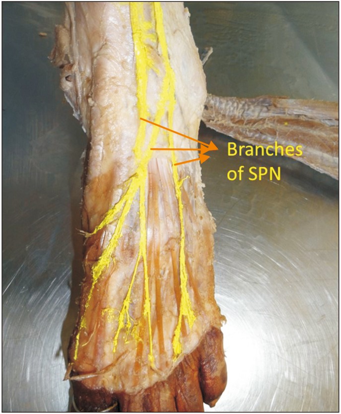
Fig. 2 In this group, first interdigital cleft is supplied by the branch of superficial peroneal nerve (SPN) instead of deep peroneal nerve (DPN). Sural nerve, lateral side of fifth toe; SPN, remaining part of the dorsum of the foot.
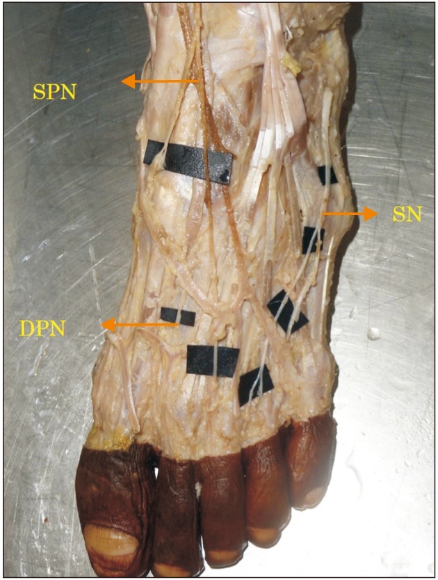
Fig. 3 In this picture lateral 2 and 1/2 finger is supplied by sural nerve (SN) instead of superficial peroneal nerve (SPN). Deep peroneal nerve (DPN), first interdigital cleft; SN, lateral 2 1/2 toes. Rest of the dorsum of the foot is by the SPN.
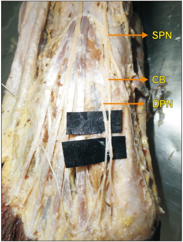
Fig. 4 Deep peroneal nerve (DPN) communicates with the medial division of superficial peroneal nerve (SPN) by communicating branch (CB) supplies first interdigital cleft. Sural nerve supplies lateral side of fifth toe. Rest of the dorsum of the foot is by the SPN.
Cited by 2 articles
-
The comprehensive review of the neurovascular supply of the ankle joint: clinical implications
Chirapat Inchai, Tanawat Vaseenon, Pasuk Mahakkanukrauh
Anat Cell Biol. 2020;53(2):126-131. doi: 10.5115/acb.20.037.The distribution pattern of the dorsal cutaneous nerves of the foot and its clinical implications
Chirapat Inchai, Tanawat Vaseenon, Yasuhito Tanaka, Pasuk Mahakkanukrauh
Anat Cell Biol. 2020;53(2):137-142. doi: 10.5115/acb.20.039.
Reference
-
1. Standring S, Borley NR, Collins P, Crossman AR, Gatzoulis MA, Healy JC, Johnson D, Mahadevan V, Newell RL, Wigley CB. Gray's anatomy: the anatomical basis of clinical practice. 40th ed. London: Churchill Livingstone;2008. p. 1427–1429.2. Tassone H, Silver MA. Superficial peroneal nerve block [Interent]. Medscape;2015. cited 2018 Oct 1. Available from: http://emedicine.medscape.com/article/83218-overview.3. Asp R, Marsland D, Elliot R. The superficial peroneal nerve: a review of its anatomy and surgical relevance. OA Anat. 2014; 2:6.4. Blair JM, Botte MJ. Surgical anatomy of the superficial peroneal nerve in the ankle and foot. Clin Orthop Relat Res. 1994; (305):229–238.5. Buntic RF, Buncke HJ, Kind GM, Chin BT, Ruebeck D, Buncke GM. The harvest and clinical application of the superficial peroneal sensory nerve for grafting motor and sensory nerve defects. Plast Reconstr Surg. 2002; 109:145–151. PMID: 11786806.6. Slutsky DJ. A practical approach to nerve grafting in the upper extremity. Atlas Hand Clin. 2005; 10:e92.7. Tallis R, Staniforth P, Fisher TR. Neurophysiological studies of autogenous sural nerve grafts. J Neurol Neurosurg Psychiatry. 1978; 41:677–683. PMID: 681954.8. Romanes GJ. Cunningham's manual of practical anatomy. 15th ed. Vol. 1. Oxford: Oxford University Press;1986. p. 16–18.9. Madhavi C, Isaac B, Antoniswamy B, Holla SJ. Anatomical variations of the cutaneous innervation patterns of the sural nerve on the dorsum of the foot. Clin Anat. 2005; 18:206–209. PMID: 15768411.10. Aktan Ikiz ZA, Uçerler H, Bilge O. The anatomic features of the sural nerve with an emphasis on its clinical importance. Foot Ankle Int. 2005; 26:560–567. PMID: 16045849.11. Drizenko A, Demondion X, Luyckx F, Mestdagh H, Cassagnaud X. The communicating branches between the sural and superficial peroneal nerves in the foot: a review of 55 cases. Surg Radiol Anat. 2004; 26:447–452. PMID: 15300414.12. Prakash , Bhardwaj AK, Singh DK, Rajini T, Jayanthi V, Singh G. Anatomic variations of superficial peroneal nerve: clinical implications of a cadaver study. Ital J Anat Embryol. 2010; 115:223–228. PMID: 21287977.13. Wahee P, Aggarwal A, Harjeet , Sahni D. Variable patterns of cutaneous innervation on the dorsum of foot in fetuses. Surg Radiol Anat. 2010; 32:469–475. PMID: 19911110.14. Gupta C, Kiruba NL, Dsouza AS, Radhakrishnan P. A morphological study to note the variable patterns of cutaneous innervation on the dorsum of foot in south Indian human foetuses and its clinical implications. Adv Biomed Res. 2013; 2:15. PMID: 23930260.15. Halm JA, Schepers T. Damage to the superficial peroneal nerve in operative treatment of fibula fractures: straight to the bone? Case report and review of the literature. J Foot Ankle Surg. 2012; 51:684–686. PMID: 22789487.
- Full Text Links
-
- Actions
-
Cited
- CITED
-
- Close
- Share
-
- Similar articles
-
- Unusual bilateral sensory innervation of the dorsum of hand by lateral antebrachial cutaneous nerve: a case report
- Anatomical variations of the innervated radial artery superficial palmar branch flap: A series of 28 clinical cases
- Uncommon configuration of intercostobrachial nerves, lateral roots, and absent medial cutaneous nerve of arm in a cadaveric study
- A Case of Cutaneous Horn Arising from Angiokeratoma in Infancy
- Neurovascular Tendocutaneous Free Flap Transplantation by Microsurgical Technique: Case Report
