Reproducibility of cone-beam computed tomographic measurements of bone plates and the interdental septum in the anterior mandible
- Affiliations
-
- 1Department of Oral Radiology, Pontifical Catholic University of Minas Gerais, Belo Horizonte, Brazil. manzi@pucminas.br
- 2Department of Dentistry, Pontifical Catholic University of Minas Gerais, Belo Horizonte, Brazil.
- KMID: 2442104
- DOI: http://doi.org/10.5624/isd.2019.49.1.9
Abstract
- PURPOSE
This study aimed to introduce a novel method to evaluate the alveolar bone and interdental septum in the anterior mandible using cone-beam computed tomography (CBCT).
MATERIALS AND METHODS
Fifty-six CBCT scans from adult patients were selected. The CBCT scans were obtained before and after orthodontic treatment. The following measurements were taken: width of the alveolar bone and the interdental septum, height of the interdental septum, height of the bone plates, distance between the cementoenamel junction and marginal bone crests, and vertical positioning of the mandibular incisor, using the lingual plane as a reference. To test the reproducibility and the stability of the lingual plane, a triangle was traced in the anterior mandible. The intra-class correlation coefficient (ICC) was used to determine intra- and inter-examiner agreement. The paired Student t-test was used to evaluate the area of the triangle and the reproducibility of all measurements.
RESULTS
The ICC was excellent for the alveolar bone and dental measurements (0.9989 and 0.9977, respectively), as well as for the interdental septum (0.9987 and 0.9961, respectively). The area of the triangles showed stability in the lingual plane (P>0.05). For the alveolar bone, mandibular incisor, and interdental septum measurements, no statistically significant differences were found between the 2 examiners (P>0.05), confirming the technical reliability of the measurements.
CONCLUSION
The method used in this study provides a valid and reproducible assessment of alveolar bone dimensions in the anterior mandible measured on CBCT images.
MeSH Terms
Figure
-

Fig. 1 On the axial slice of the mandible, the localization of the parasagittal slices was determined for the measurements of the alveolar bone (continuous AB lines) and the measurements of the interdental septum (interrupted CD lines).
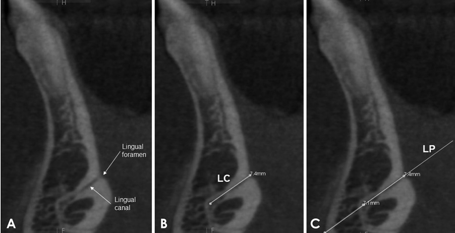
Fig. 2 Parasagittal slice. A. Localization of the lingual foramen and the mandibular lingual canal. B. Tracing of the mandibular lingual canal (LC). C. Extension of the lingual canal line to obtain the lingual plane (LP).
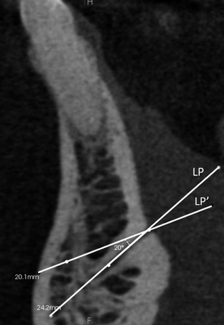
Fig. 3 Parasagittal slice. Tracing of the compensatory lingual plane (LP′), with 20° of compensation. LP: lingual plane.
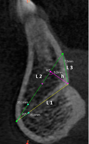
Fig. 4 Parasagittal slice. Tracing of the triangle. First, line 1 was traced over the lingual plane, respecting the limits of the symphysis (L1). The second side was traced with an internal angle of 20°, beginning from the facial limit of L1 (L2). The third side joined the lingual extremities of sides L1 and L2, forming L3. Height of the triangle (h).
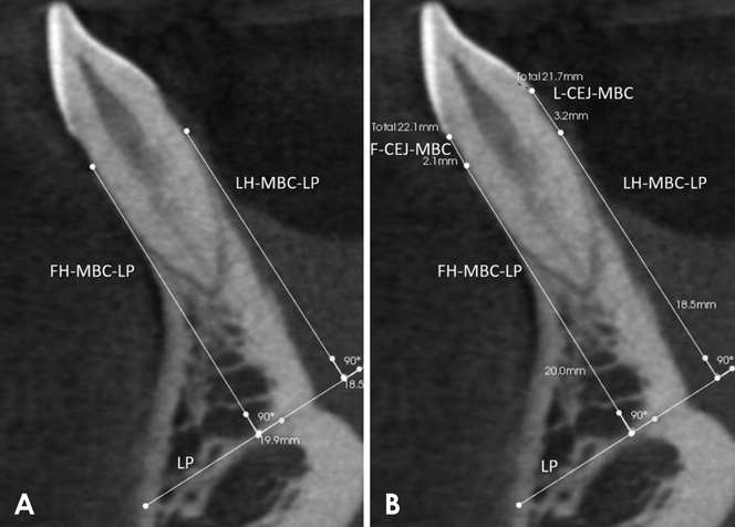
Fig. 5 Parasagittal slice. A. Measurement of the height of the facial bone plate (FH-MBC-LP) and the lingual bone plate (LH-MBC-LP). B. Measurement of the distance between the CEJ and the marginal bone crest on the facial side (F-CEJ-MBC) and on the lingual side (L-CEJ-MBC). MBC: marginal bone crest; LP: lingual plane.
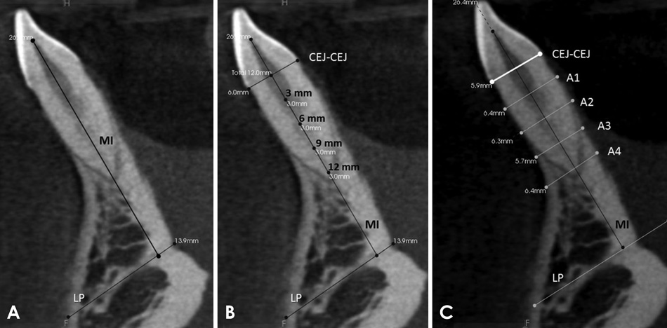
Fig. 6 Parasagittal slice. A. Measurement of the vertical positioning of the mandibular incisor (MI line). B and C. Measurement of the thickness of the alveolar bone. Initially, a line was traced along the incisor's long axis (MI line) and the joining line between the facial and lingual CEJs (CEJ-CEJ line). Next, the thicknesses of the alveolar process were measured using the A1, A2, A3, and A4 lines, traced 3 mm, 6 mm, 9 mm, and 12 mm below the CEJ-CEJ line, respectively.

Fig. 7 Parasagittal slice. A. Measurement of the height of the interdental septum through the SH line, traced perpendicular to the lingual plane (LP). B. Measurement of the thickness of the interdental septum at points located 3 mm (S1), 6 mm (S2), 9 mm (S3), and 12 mm (S4) below the bone crest. LP: lingual plane, SH: bone crest height.

Fig. 8 Triangle areas obtained at time 1 (T1) and time 2 (T2) (NS: P>0.05).
Reference
-
1. Garlock DT, Buschang PH, Araujo EA, Behrents RG, Kim KB. Evaluation of marginal alveolar bone in the anterior mandible with pretreatment and posttreatment computed tomography in nonextraction patients. Am J Orthod Dentofacial Orthop. 2016; 149:192–201.
Article2. Sarikaya S, Haydar B, Ciğer S, Ariyürek M. Changes in alveolar bone thickness due to retraction of anterior teeth. Am J Orthod Dentofacial Orthop. 2002; 122:15–26.
Article3. Fuhrmann R. Three-dimensional interpretation of labiolingual bone width of the lower incisors. Part II. J Orofac Orthop. 1996; 57:168–185.4. Pini-Prato G, Nieri M, Pagliaro U, Giorgi TS, La Marca M, Franceschi D, et al. Surgical treatment of single gingival recessions: clinical guidelines. Eur J Oral Implantol. 2014; 7:9–43.5. Fuhrmann RA, Wehrbein H, Langen HJ, Diedrich PR. Assessment of the dentate alveolar process with high resolution computed tomography. Dentomaxillofac Radiol. 1995; 24:50–54.
Article6. Merli M, Merli I, Raffaelli E, Pagliaro U, Nastri L, Nieri M. Bone augmentation at implant dehiscences and fenestrations. A systematic review of randomised controlled trials. Eur J Oral Implantol. 2016; 9:11–32.7. Takeshita WM, Vessoni Iwaki LC, Da Silva MC, Tonin RH. Evaluation of diagnostic accuracy of conventional and digital periapical radiography, panoramic radiography, and cone-beam computed tomography in the assessment of alveolar bone loss. Contemp Clin Dent. 2014; 5:318–323.
Article8. Kumar V, Ludlow J, Soares Cevidanes LH, Mol A. In vivo comparison of conventional and cone beam CT synthesized cephalograms. Angle Orthod. 2008; 78:873–879.
Article9. Mozzo P, Procacci C, Tacconi A, Martini PT, Andreis IA. A new volumetric CT machine for dental imaging based on the cone-beam technique: preliminary results. Eur Radiol. 1998; 8:1558–1564.
Article10. Babiuc I, Tărlungeanu I, Păuna M. Cone beam computed tomography observations of the lingual foramina and their bony canals in the median region of the mandible. Rom J Morphol Embryol. 2011; 52:827–829.11. Shrout PE, Fleiss JL. Intraclass correlations: uses in assessing rater reliability. Psychol Bull. 1979; 86:420–428.
Article12. Dahlberg G. Standard error and medicine. Acta Genet Stat Med. 1949; 1:313–321.
Article13. Begley CG, Ioannidis JP. Reproducibility in science: improving the standard for basic and preclinical research. Circ Res. 2015; 116:116–126.14. Bimstein E, Crevoisier RA, King DL. Changes in the morphology of the buccal alveolar bone of protruded mandibular permanent incisors secondary to orthodontic alignment. Am J Orthod Dentofacial Orthop. 1990; 97:427–430.15. Qu X, Liu Z, Wang Y, Fang Y, Du M, He H. Dentofacial traits in association with lower incisor alveolar cancellous bone thickness: a multiple regression analysis. Angle Orthod. 2017; 87:409–415.
Article16. Baysal A, Uysal T, Veli I, Ozer T, Karadede I, Hekimoglu S. Evaluation of alveolar bone loss following rapid maxillary expansion using cone-beam computed tomography. Korean J Orthod. 2013; 43:83–95.
Article17. Garib DG, Menezes MH, Silva Filho OG, Santos PB. Immediate periodontal bone plate changes induced by rapid maxillary expansion in the early mixed dentition: CT findings. Dental Press J Orthod. 2014; 19:36–43.
Article18. Dudic A, Giannopoulou C, Leuzinger M, Kiliaridis S. Detection of apical root resorption after orthodontic treatment by using panoramic radiography and cone-beam computed tomography of super-high resolution. Am J Orthod Dentofacial Orthop. 2009; 135:434–437.
Article19. Yared KF, Zenobio EG, Pacheco W. Periodontal status of mandibular central incisors after orthodontic proclination in adults. Am J Orthod Dentofacial Orthop. 2006; 130:6.e1–6.e8.
Article20. Buschang PH, Julien K, Sachdeva R, Demirjian A. Childhood and pubertal growth changes of the human symphysis. Angle Orthod. 1992; 62:203–210.21. Aki T, Nanda RS, Currier GF, Nanda SK. Assessment of symphysis morphology as a predictor of the direction of mandibular growth. Am J Orthod Dentofacial Orthop. 1994; 106:60–69.22. Sun Z, Smith T, Kortam S, Kim DG, Tee BC, Fields H. Effect of bone thickness on alveolar bone-height measurements from cone-beam computed tomography images. Am J Orthod Dentofacial Orthop. 2011; 139:e117–e127.
Article23. Timock AM, Cook V, McDonald T, Leo MC, Crowe J, Benninger BL, et al. Accuracy and reliability of buccal bone height and thickness measurements from cone-beam computed tomography imaging. Am J Orthod Dentofacial Orthop. 2011; 140:734–744.
Article24. Leung CC, Palomo L, Griffith R, Hans MG. Accuracy and reliability of cone-beam computed tomography for measuring alveolar bone height and detecting bony dehiscences and fenestrations. Am J Orthod Dentofacial Orthop. 2010; 137:S109–S119.
Article25. Lund H, Gröndahl K, Gröndahl HG. Cone beam computed tomography evaluations of marginal alveolar bone before and after orthodontic treatment combined with premolar extractions. Eur J Oral Sci. 2012; 120:201–211.
Article
- Full Text Links
-
- Actions
-
Cited
- CITED
-
- Close
- Share
-
- Similar articles
-
- Central odontogenic fibroma (simple type) in a four-year-old boy: atypical cone-beam computed tomographic appearance with periosteal reaction
- Buccal cortical bone thickness on CBCT for mini-implant
- Evaluation of interdental distance of natural teeth with cone-beam computerized tomography
- Bone changes in the mandibular incisors after orthodontic correction of dental crowding without extraction: A cone-beam computed tomographic evaluation
- Three-dimensional evaluation of maxillary anterior alveolar bone for optimal placement of miniscrew implants
