J Breast Cancer.
2019 Mar;22(1):77-85. 10.4048/jbc.2019.22.e9.
CD9 Expression in Tumor Cells Is Associated with Poor Prognosis in Patients with Invasive Lobular Carcinoma
- Affiliations
-
- 1Department of Pathology, Yeungnam University College of Medicine, Daegu, Korea. ykbae@ynu.ac.kr
- 2Department of Breast Surgery, Yeungnam University College of Medicine, Daegu, Korea.
- 3Department of Biochemistry and Molecular Biology, Yeungnam University College of Medicine, Daegu, Korea.
- KMID: 2441853
- DOI: http://doi.org/10.4048/jbc.2019.22.e9
Abstract
- PURPOSE
We investigated the prognostic significance of CD9 expression in tumor cells of patients with invasive lobular carcinoma (ILC).
METHODS
CD9 expression was evaluated by immunohistochemistry in 113 ILC tissue samples. Correlation of CD9 expression with the patients' clinicopathological parameters and overall survival was assessed.
RESULTS
CD9 expression was detected in 48 (42.5%) ILC patients. However, no significant relation could be determined between CD9 expression and the clinicopathological parameters of the patient including tumor size, lymph node metastasis, lymphovascular invasion, histologic grade, expression of hormone receptors, human epidermal growth factor receptor 2 status, and Ki-67 labeling index. Patients with CD9 expression had worse overall survival (p = 0.051) and disease-free survival (DFS, p = 0.014) compared to patients without CD9 expression. Multivariate analysis revealed that CD9 expression was an independent prognostic factor for DFS (p = 0.049).
CONCLUSION
CD9 expression in tumor cells could be a significant prognostic marker in patients with ILC.
MeSH Terms
Figure
-
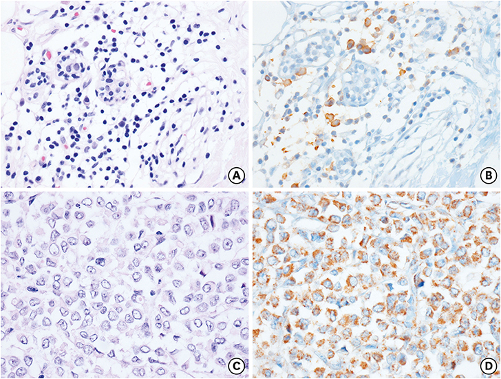
Figure 1 Representative immunohistochemical results for CD9 expression in invasive lobular carcinoma. (A, B) Negative CD9 expression in non-neoplastic mammary glands and positive CD9 expression in plasma cells infiltrated within lobules. (C, D) Diffuse and strong CD9 expression in tumor cells. A and C, hematoxylin and eosin stain; B and D, immunohistochemistry for CD9; magnification of all figures, ×400.
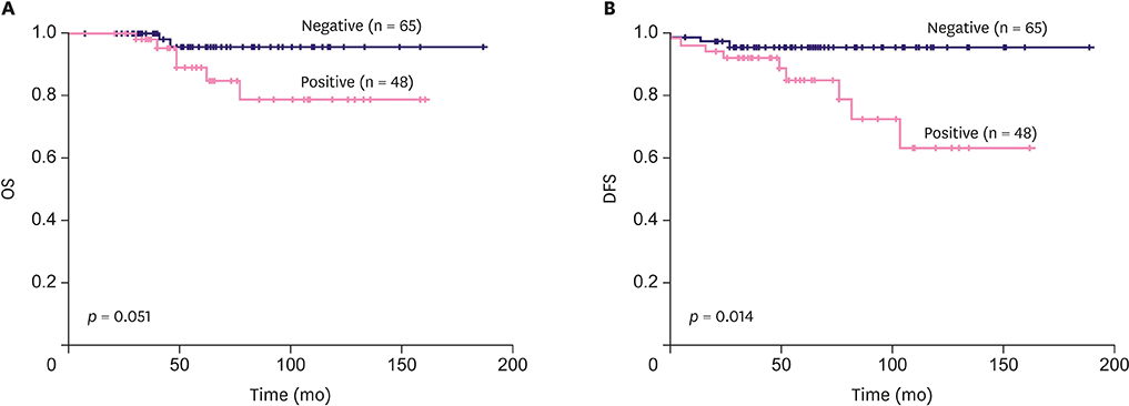
Figure 2 Kaplan-Meier survival curves according to CD9 expression in patients with invasive lobular carcinoma. (A) OS. (B) DFS. OS = overall survival; DFS = disease-free survival.
Reference
-
1. Lazo PA. Functional implications of tetraspanin proteins in cancer biology. Cancer Sci. 2007; 98:1666–1677.
Article2. Stipp CS, Kolesnikova TV, Hemler ME. Functional domains in tetraspanin proteins. Trends Biochem Sci. 2003; 28:106–112.
Article3. Huang C, Taki T, Adachi M, Yagita M, Sawada S, Takabayashi A, et al. MRP-1/CD9 and KAI1/CD82 expression in normal and various cancer tissues. Int J Oncol. 1997; 11:1045–1051.
Article4. Miyake M, Nakano K, Itoi SI, Koh T, Taki T. Motility-related protein-1 (MRP-1/CD9) reduction as a factor of poor prognosis in breast cancer. Cancer Res. 1996; 56:1244–1249.5. Huang CI, Kohno N, Ogawa E, Adachi M, Taki T, Miyake M. Correlation of reduction in MRP-1/CD9 and KAI1/CD82 expression with recurrences in breast cancer patients. Am J Pathol. 1998; 153:973–983.
Article6. Uchida S, Shimada Y, Watanabe G, Li ZG, Hong T, Miyake M, et al. Motility-related protein (MRP-1/CD9) and KAI1/CD82 expression inversely correlate with lymph node metastasis in oesophageal squamous cell carcinoma. Br J Cancer. 1999; 79:1168–1173.
Article7. Kusukawa J, Ryu F, Kameyama T, Mekada E. Reduced expression of CD9 in oral squamous cell carcinoma: CD9 expression inversely related to high prevalence of lymph node metastasis. J Oral Pathol Med. 2001; 30:73–79.
Article8. Buim ME, Lourenço SV, Carvalho KC, Cardim R, Pereira C, Carvalho AL, et al. Downregulation of CD9 protein expression is associated with aggressive behavior of oral squamous cell carcinoma. Oral Oncol. 2010; 46:166–171.
Article9. Hori H, Yano S, Koufuji K, Takeda J, Shirouzu K. CD9 expression in gastric cancer and its significance. J Surg Res. 2004; 117:208–215.
Article10. Soyuer S, Soyuer I, Unal D, Ucar K, Yildiz OG, Orhan O. Prognostic significance of CD9 expression in locally advanced gastric cancer treated with surgery and adjuvant chemoradiotherapy. Pathol Res Pract. 2010; 206:607–610.
Article11. Huan J, Gao Y, Xu J, Sheng W, Zhu W, Zhang S, et al. Overexpression of CD9 correlates with tumor stage and lymph node metastasis in esophageal squamous cell carcinoma. Int J Clin Exp Pathol. 2015; 8:3054–3061.12. Kwon HJ, Min SY, Park MJ, Lee C, Park JH, Chae JY, et al. Expression of CD9 and CD82 in clear cell renal cell carcinoma and its clinical significance. Pathol Res Pract. 2014; 210:285–290.
Article13. Kwon HJ, Choi JE, Kang SH, Son Y, Bae YK. Prognostic significance of CD9 expression differs between tumour cells and stromal immune cells, and depends on the molecular subtype of the invasive breast carcinoma. Histopathology. 2017; 70:1155–1165.
Article14. Lakhani SR, Rakha E, Simpson PT. Invasive lobular carcinoma. In : Lakhani SR, Ellis IO, Schnitt SJ, Tan PH, van de Vijver MJ, editors. WHO Classification of Tumors of the Breast. 4th ed. Lyon: IARC;2012. p. 40–42.15. Lee JH, Park S, Park HS, Park BW. Clinicopathological features of infiltrating lobular carcinomas comparing with infiltrating ductal carcinomas: a case control study. World J Surg Oncol. 2010; 8:34.
Article16. Park JS, Choi DH, Huh SJ, Park W, Kim YI, Nam SJ, et al. Comparison of clinicopathological features and treatment results between invasive lobular carcinoma and ductal carcinoma of the breast. J Breast Cancer. 2015; 18:285–290.
Article17. Hammond ME, Hayes DF, Dowsett M, Allred DC, Hagerty KL, Badve S, et al. American Society of Clinical Oncology/College of American Pathologists guideline recommendations for immunohistochemical testing of estrogen and progesterone receptors in breast cancer. J Clin Oncol. 2010; 28:2784–2795.
Article18. Wolff AC, Hammond ME, Hicks DG, Dowsett M, McShane LM, Allison KH, et al. Recommendations for human epidermal growth factor receptor 2 testing in breast cancer: American Society of Clinical Oncology/College of American Pathologists clinical practice guideline update. J Clin Oncol. 2013; 31:3997–4013.
Article19. Zöller M. Tetraspanins: push and pull in suppressing and promoting metastasis. Nat Rev Cancer. 2009; 9:40–55.
Article20. Shi W, Fan H, Shum L, Derynck R. The tetraspanin CD9 associates with transmembrane TGF-alpha and regulates TGF-alpha-induced EGF receptor activation and cell proliferation. J Cell Biol. 2000; 148:591–602.
Article21. Rappa G, Green TM, Karbanová J, Corbeil D, Lorico A. Tetraspanin CD9 determines invasiveness and tumorigenicity of human breast cancer cells. Oncotarget. 2015; 6:7970–7991.
Article22. Kischel P, Bellahcene A, Deux B, Lamour V, Dobson R, DE Pauw E, et al. Overexpression of CD9 in human breast cancer cells promotes the development of bone metastases. Anticancer Res. 2012; 32:5211–5220.23. Longo N, Yáñez-Mó M, Mittelbrunn M, de la Rosa G, Muñoz ML, Sánchez-Madrid F, et al. Regulatory role of tetraspanin CD9 in tumor-endothelial cell interaction during transendothelial invasion of melanoma cells. Blood. 2001; 98:3717–3726.
Article24. Kohmo S, Kijima T, Otani Y, Mori M, Minami T, Takahashi R, et al. Cell surface tetraspanin CD9 mediates chemoresistance in small cell lung cancer. Cancer Res. 2010; 70:8025–8035.
Article
- Full Text Links
-
- Actions
-
Cited
- CITED
-
- Close
- Share
-
- Similar articles
-
- CD9 Expression in Colorectal Carcinomas and Its Prognostic Significance
- Nodular Metastatic Carcinoma from Invasive Lobular Breast Cancer
- Invasive Lobular Carcinoma of the Breast Associated with Mixed Lobular and Ductal Carcinoma In Situ: A Case Report
- Expression and Clinicopathological Significance of CD9 in Gastrointestinal Stromal Tumor
- Multi-Focal Lobular Carcinoma In Situ Arising in Benign Phyllodes Tumor: A Case Report
