Tumor-Associated Macrophages as Potential Prognostic Biomarkers of Invasive Breast Cancer
- Affiliations
-
- 1Department of Pathology, Keimyung University School of Medicine, Daegu, Korea. pathol72@gmail.com
- 2Department of General Surgery, Keimyung University School of Medicine, Daegu, Korea.
- 3Department of Pathology, Samsung Medical Center, Sungkyunkwan University School of Medicine, Seoul, Korea.
- KMID: 2441850
- DOI: http://doi.org/10.4048/jbc.2019.22.e5
Abstract
- PURPOSE
Tumor-associated macrophages (TAMs) are activated macrophages associated with tumor progression in various cancers. TAMs can polarize M1 or M2 type. M1 has a pro-inflammatory function and kills pathogens. Conversely, M2 shows immunosuppressive action and promotes tumor growth. There are various markers of TAMs. CD11c is considered as a specific marker of M1. CD163 is an optimal marker for M2. CD68 is known as a pan-macrophage marker. We evaluated the relationship between the clinicopathological parameters and immunohistochemical expressions of CD11c, CD163, and CD68 in invasive breast cancer (IBC), and the prognostic value of macrophage localization within the tumor stroma (TS) and tumor nest (TN).
METHODS
Immunohistochemistry of CD68, CD11c, and CD163 was analyzed on tissue microarrays of 367 IBCs. The number of CD68+, CD11c+, or CD163+ macrophages in TN vs. TS was counted by 2 pathologists. The correlations between the degree of macrophage (CD68+, CD11c+, or CD163+) infiltration and the clinicopathological parameters were analyzed. We also assessed the impact of macrophages (CD68+, CD11c+, or CD163+) on disease free survival (DFS) and overall survival (OS).
RESULTS
High numbers of macrophages (CD68+, CD11c+, or CD163+) were associated with higher histologic grade, higher Ki-67 proliferating index, estrogen receptor negativity, and progesterone receptor negativity. High numbers of macrophages (CD11c+ or CD163+) in TS were associated with a larger tumor size. Furthermore, CD163+ macrophages in TN were an independent prognostic marker of reduced OS and DFS. Conversely, CD11c+ macrophages in TS were an independent prognostic marker for higher OS and DFS.
CONCLUSION
TAMs, including M2 type, are associated with tumor progression in IBC. They can also act as a significant unfavorable or favorable prognostic factor. In addition to simply analyzing the degree of TAM infiltration, it is also important to analyze the location of TAMs.
MeSH Terms
Figure
-

Figure 1 Representative images of IHC analysis of TAM markers in IBC. (A) CD68+ macrophages are mostly present in TS (IHC for CD68, 400× magnification). (B) CD68+ macrophages are present in both TS and TN (IHC for CD68, 400× magnification). (C) CD11c+ macrophages are mainly present in TS (IHC for CD11c, 400× magnification). (D) CD11c+ macrophages are mostly present in TN (IHC for CD11c, 400× magnification). (E) CD163+ macrophages are chiefly present in TS (IHC for CD163, 400× magnification). (F) CD163+ macrophages are present in both TS and TN (IHC for CD163, 400× magnification). IBC = invasive breast cancer; IHC = immunohistochemistry; TAM = tumor-associated macrophage; TS = tumor stroma; TN = tumor nest.
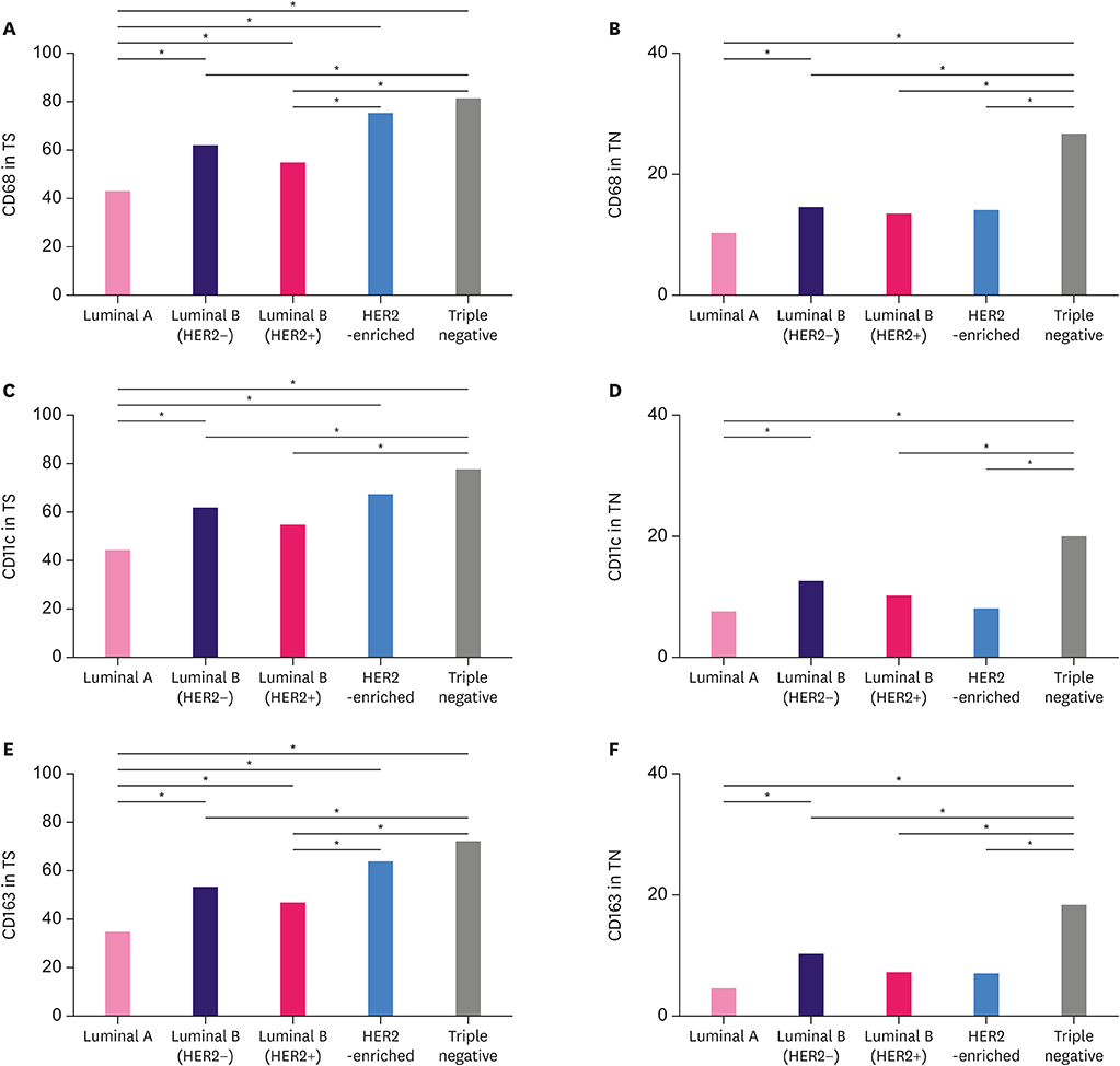
Figure 2 Mean of the macrophages (CD68+, CD11c+, or CD163+) according to each molecular subtype. (A) CD68 in TS, (B) CD68 in TN, (C) CD11c in TS, (D) CD11c in TN, (E) CD163 in TS, and (F) CD163 in TN. HER2 = human epithelial growth factor receptor 2; TS = tumor stroma; TN = tumor nest. *p<0.05.
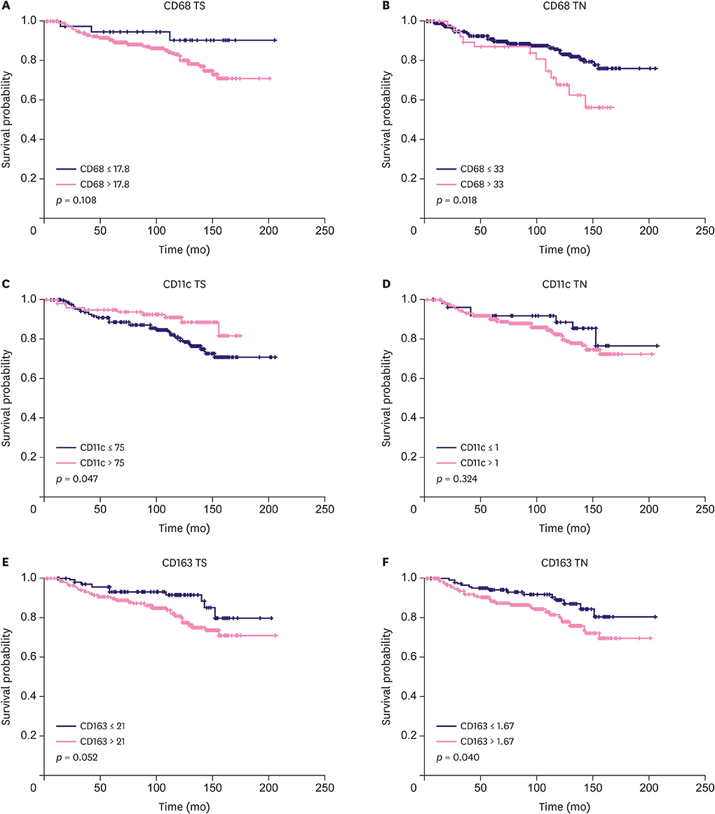
Figure 3 The OS curves according to the infiltration density of CD68+ macrophages in TS (A) and TN (B), CD11+ macrophages in TS (C) and TN (D), and CD163+ macrophages in TS (E) and TN (F) in IBC. OS = overall survival; TS = tumor stroma; TN = tumor nest; IBC = invasive breast cancer.
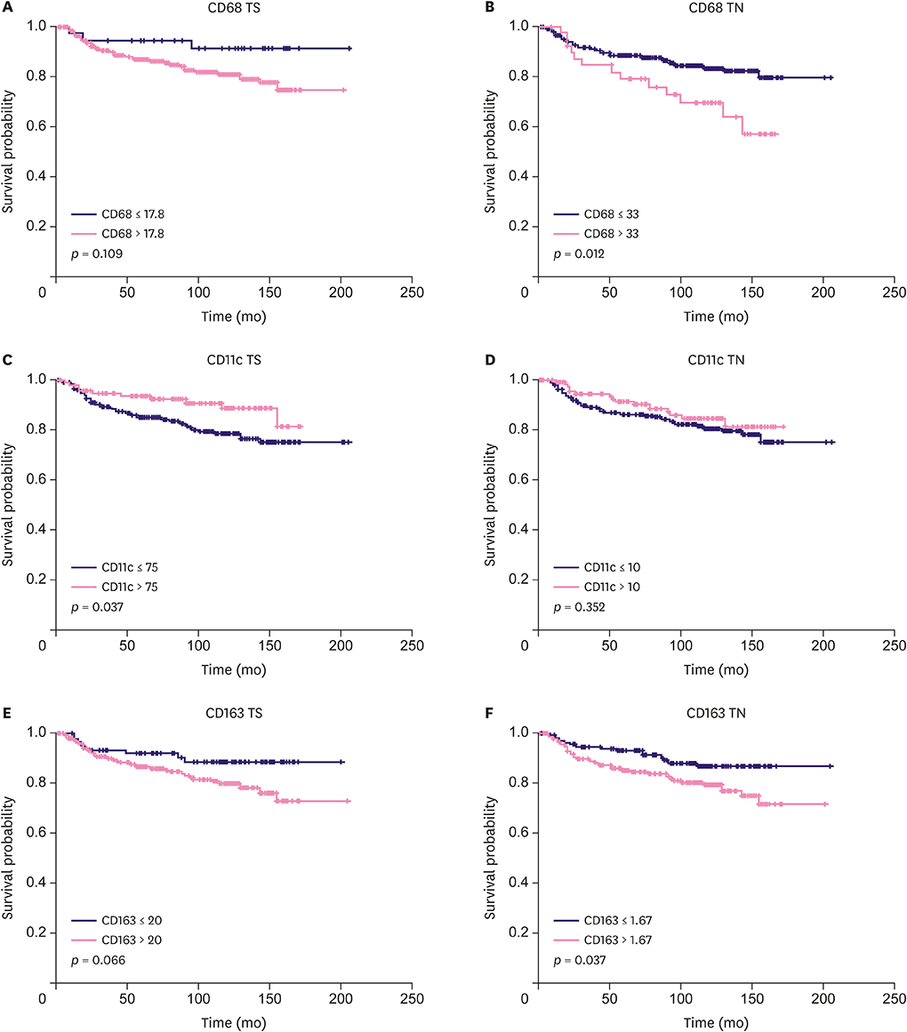
Figure 4 Disease free curves based on the infiltration density of CD68+ macrophages in TS (A) and TN (B), CD11+ macrophages in TS (C) and TN (D), and CD163+ macrophages in TS (E) and TN (F) in IBC. TS = tumor stroma; TN = tumor nest; IBC = invasive breast cancer.
Reference
-
1. Burugu S, Asleh-Aburaya K, Nielsen TO. Immune infiltrates in the breast cancer microenvironment: detection, characterization and clinical implication. Breast Cancer. 2017; 24:3–15.
Article2. Mahmoud SM, Lee AH, Paish EC, Macmillan RD, Ellis IO, Green AR. Tumour-infiltrating macrophages and clinical outcome in breast cancer. J Clin Pathol. 2012; 65:159–163.
Article3. Medrek C, Pontén F, Jirström K, Leandersson K. The presence of tumor associated macrophages in tumor stroma as a prognostic marker for breast cancer patients. BMC Cancer. 2012; 12:306.
Article4. Gwak JM, Jang MH, Kim DI, Seo AN, Park SY. Prognostic value of tumor-associated macrophages according to histologic locations and hormone receptor status in breast cancer. PLoS One. 2015; 10:e0125728.
Article5. Yang J, Li X, Liu X, Liu Y. The role of tumor-associated macrophages in breast carcinoma invasion and metastasis. Int J Clin Exp Pathol. 2015; 8:6656–6664.6. Lindsten T, Hedbrant A, Ramberg A, Wijkander J, Solterbeck A, Eriksson M, et al. Effect of macrophages on breast cancer cell proliferation, and on expression of hormone receptors, uPAR and HER-2. Int J Oncol. 2017; 51:104–114.
Article7. Gordon S, Martinez FO. Alternative activation of macrophages: mechanism and functions. Immunity. 2010; 32:593–604.
Article8. Mantovani A, Sozzani S, Locati M, Allavena P, Sica A. Macrophage polarization: tumor-associated macrophages as a paradigm for polarized M2 mononuclear phagocytes. Trends Immunol. 2002; 23:549–555.
Article9. Nizet V, Johnson RS. Interdependence of hypoxic and innate immune responses. Nat Rev Immunol. 2009; 9:609–617.
Article10. Gordon S. Alternative activation of macrophages. Nat Rev Immunol. 2003; 3:23–35.
Article11. Wynn TA. Fibrotic disease and the T(H)1/T(H)2 paradigm. Nat Rev Immunol. 2004; 4:583–594.
Article12. Weber M, Büttner-Herold M, Hyckel P, Moebius P, Distel L, Ries J, et al. Small oral squamous cell carcinomas with nodal lymphogenic metastasis show increased infiltration of M2 polarized macrophages--an immunohistochemical analysis. J Craniomaxillofac Surg. 2014; 42:1087–1094.
Article13. Ambarus CA, Krausz S, van Eijk M, Hamann J, Radstake TR, Reedquist KA, et al. Systematic validation of specific phenotypic markers for in vitro polarized human macrophages. J Immunol Methods. 2012; 375:196–206.
Article14. Liu C, Li Y, Yu J, Feng L, Hou S, Liu Y, et al. Targeting the shift from M1 to M2 macrophages in experimental autoimmune encephalomyelitis mice treated with fasudil. PLoS One. 2013; 8:e54841.
Article15. Edge SB, Byrd DR, Compton CC, Fritz AG, Greene FL, Trotti A. AJCC Cancer Staging Manual. New York (NY): Springer;2010.16. Lakhani SR, Ellis IO, Schnitt SJ, Tan PH, van de Vijver MJ. WHO Classification of Tumours of the Breast. Lyon: International Agency for Research on Cancer;2012.17. Hammond ME, Hayes DF, Dowsett M, Allred DC, Hagerty KL, Badve S, et al. American Society of Clinical Oncology/College of American Pathologists guideline recommendations for immunohistochemical testing of estrogen and progesterone receptors in breast cancer. Arch Pathol Lab Med. 2010; 134:907–922.
Article18. Wolff AC, Hammond ME, Hicks DG, Dowsett M, McShane LM, Allison KH, et al. Recommendations for human epidermal growth factor receptor 2 testing in breast cancer: American Society of Clinical Oncology/College of American Pathologists clinical practice guideline update. J Clin Oncol. 2013; 31:3997–4013.
Article19. Dowsett M, Nielsen TO, A'Hern R, Bartlett J, Coombes RC, Cuzick J, et al. Assessment of Ki67 in breast cancer: recommendations from the International Ki67 in Breast Cancer working group. J Natl Cancer Inst. 2011; 103:1656–1664.
Article20. Guiu S, Michiels S, André F, Cortes J, Denkert C, Di Leo A, et al. Molecular subclasses of breast cancer: how do we define them? The IMPAKT 2012 working group statement. Ann Oncol. 2012; 23:2997–3006.
Article21. Zhang M, Sun H, Zhao S, Wang Y, Pu H, Wang Y, et al. Expression of PD-L1 and prognosis in breast cancer: a meta-analysis. Oncotarget. 2017; 8:31347–31354.
Article22. Bae SB, Cho HD, Oh MH, Lee JH, Jang SH, Hong SA, et al. Expression of programmed death receptor ligand 1 with high tumor-infiltrating lymphocytes is associated with better prognosis in breast cancer. J Breast Cancer. 2016; 19:242–251.
Article23. Santoni M, Romagnoli E, Saladino T, Foghini L, Guarino S, Capponi M, et al. Triple negative breast cancer: key role of tumor-associated macrophages in regulating the activity of anti-PD-1/PD-L1 agents. Biochim Biophys Acta Rev Cancer. 2018; 1869:78–84.
Article24. Bingle L, Brown NJ, Lewis CE. The role of tumour-associated macrophages in tumour progression: implications for new anticancer therapies. J Pathol. 2002; 196:254–265.
Article25. Sousa S, Brion R, Lintunen M, Kronqvist P, Sandholm J, Mönkkönen J, et al. Human breast cancer cells educate macrophages toward the M2 activation status. Breast Cancer Res. 2015; 17:101.
Article26. Kurahara H, Shinchi H, Mataki Y, Maemura K, Noma H, Kubo F, et al. Significance of M2-polarized tumor-associated macrophage in pancreatic cancer. J Surg Res. 2011; 167:e211–e219.
Article27. Wang Y, Xu B, Hu WW, Chen LJ, Wu CP, Lu BF, et al. High expression of CD11c indicates favorable prognosis in patients with gastric cancer. World J Gastroenterol. 2015; 21:9403–9412.
Article28. Castro FV, Tutt AL, White AL, Teeling JL, James S, French RR, et al. CD11c provides an effective immunotarget for the generation of both CD4 and CD8 T cell responses. Eur J Immunol. 2008; 38:2263–2273.
Article29. Jensen TO, Schmidt H, Møller HJ, Høyer M, Maniecki MB, Sjoegren P, et al. Macrophage markers in serum and tumor have prognostic impact in American Joint Committee on Cancer stage I/II melanoma. J Clin Oncol. 2009; 27:3330–3337.
Article30. Ohno S, Ohno Y, Suzuki N, Kamei T, Koike K, Inagawa H, et al. Correlation of histological localization of tumor-associated macrophages with clinicopathological features in endometrial cancer. Anticancer Res. 2004; 24:3335–3342.
- Full Text Links
-
- Actions
-
Cited
- CITED
-
- Close
- Share
-
- Similar articles
-
- Prognostic Impact and Clinicopathological Correlation of CD133 and ALDH1 Expression in Invasive Breast Cancer
- Matrix Metallopeptidase 3 Polymorphisms: Emerging genetic Markers in Human Breast Cancer Metastasis
- Introduction of a New Staging System of Breast Cancer for Radiologists: An Emphasis on the Prognostic Stage
- Downregulation of N-myc and STAT Interactor Protein Predicts Aggressive Tumor Behavior and Poor Prognosis in Invasive Ductal Carcinoma
- Application of Biomarkers for the Prediction and Diagnosis of Bone Metastasis in Breast Cancer
