Change of Extracellular Glutamate Level in Striatum during Deep Brain Stimulation of the Entopeduncular Nucleus in Rats
- Affiliations
-
- 1Department of Science in Korean Medicine, Graduate School, College of Korean Medicine, Kyung Hee University, Seoul, Korea.
- 2Department of Neurosurgery, St. Vincent's Hospital, College of Medicine, The Catholic University of Korea, Seoul, Korea. medics0919@gmail.com
- KMID: 2441560
- DOI: http://doi.org/10.3340/jkns.2018.0122
Abstract
OBJECTIVE
Globus pallidus interna (GPi) is acknowledged as an essential treatment for advanced Parkinson's disease (PD). Nonetheless, the neurotransmitter study about its results is undiscovered. The goal of this research was to examine influences of entopeduncular nucleus (EPN) stimulation, identical to human GPi, in no-lesioned (NL) rat and 6-hydroxydopamine (6-HD)-lesioned rat on glutamate change in the striatum.
METHODS
Extracellular glutamate level changes in striatum of NL category, NL with deep brain stimulation (DBS) category, 6-HD category, and 6-HD with DBS category were examined using microdialysis and high-pressure liquid chromatography. Tyrosine hydroxylase (TH) immunoreactivities in substantia nigra and striatum of the four categories were also analyzed.
RESULTS
Extracellular glutamate levels in the striatum of NL with DBS category and 6-HD with DBS category were significantly increased by EPN stimulation compared to those in the NL category and 6-HD category. EPN stimulation had no significant effect on the expression of TH in NL or 6-HD category.
CONCLUSION
Clinical results of GPi DBS are not only limited to direct inhibitory outflow to thalamus. They also include extensive alteration within basal ganglia.
MeSH Terms
-
Animals
Basal Ganglia
Chromatography, Liquid
Deep Brain Stimulation*
Entopeduncular Nucleus*
Globus Pallidus
Glutamates
Glutamic Acid*
Humans
Microdialysis
Neurotransmitter Agents
Oxidopamine
Parkinson Disease
Rats*
Substantia Nigra
Thalamus
Tyrosine 3-Monooxygenase
Glutamates
Glutamic Acid
Neurotransmitter Agents
Oxidopamine
Tyrosine 3-Monooxygenase
Figure
-
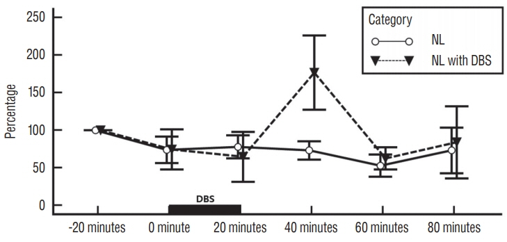
Fig. 1. Influence of EPN stimulation on striatal glutamate levels in no-lesioned rats. Each point represents the mean±standard error from six rats per category : no-lesioned rats without deep brain stimulation (NL category) and no-lesioned rats with deep brain stimulation (NL with DBS category). DBS : deep brain stimulation, NL : no-lesioned, EPN : entopeduncular nucleus.
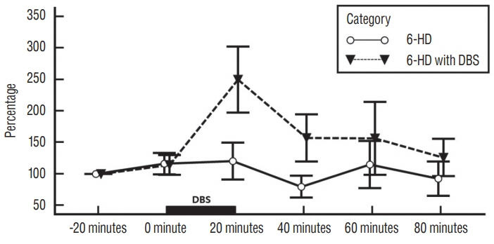
Fig. 2. Influence of EPN stimulation on striatal glutamate levels in 6-HD lesioned rats. Each point represents the mean±standard error from six rats per category : 6-HD lesioned rats without DBS (6-HD category) and 6-HD lesioned rats with deep brain stimulation (6-HD with DBS category). DBS : deep brain stimulation, 6-HD : 6-hydroxydopamine, EPN : entopeduncular nucleus.
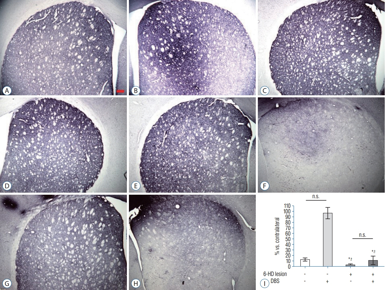
Fig. 3. TH-immunoreactivity in the Striatum. Representative images showed TH-immunoreactivity in the striatum of the ipsilateral (A, C, E, G) and contralateral (B, D, F, H) sides. Scale bar=200 μm. A and B : NL category. C and D : NL with DBS category. E and F : 6-HD category. G and H : 6-HD with DBS category. Mean (±standard error of mean) quantification (I) of TH expression showed significant decrease in both 6-HD and 6-HD with DBS categories compared to NL categories (n=5–6 per groups). *p<0.001 versus NL category. † p<0.001 versus NL with DBS category, ANOVA. n.s. : no significance, 6-HD : 6-hydroxydopamine, DBS : deep brain stimulation, TH : tyrosine hydroxylase, NL : no-lesioned, ANOVA : analysis of variance.

Fig. 4. TH-immunoreactivity in the SN. Representative images showed TH-immunoreactivity in the SN. Left side is ipsilateral part and right side is contralateral part of the SN. Scale bar=200 μm. A : NL category. B : NL with DBS category. C : 6-HD category. D : 6-HD with DBS category. Mean (±standard error of mean) quantification (E) of TH expression showed significant decrease in both 6-HD category and 6-HD with DBS category compared to NL categories (n=4–6 per groups). *p<0.01 and † p <0.001 versus NL category. ‡ p<0.001 versus NL with DBS category, ANOVA. n.s. : no significance, 6-HD : 6-hydroxydopamine, DBS : deep brain stimulation, SN : substantia nigra, TH : tyrosine hydroxylase, NL : no-lesioned, ANOVA : analysis of variance.
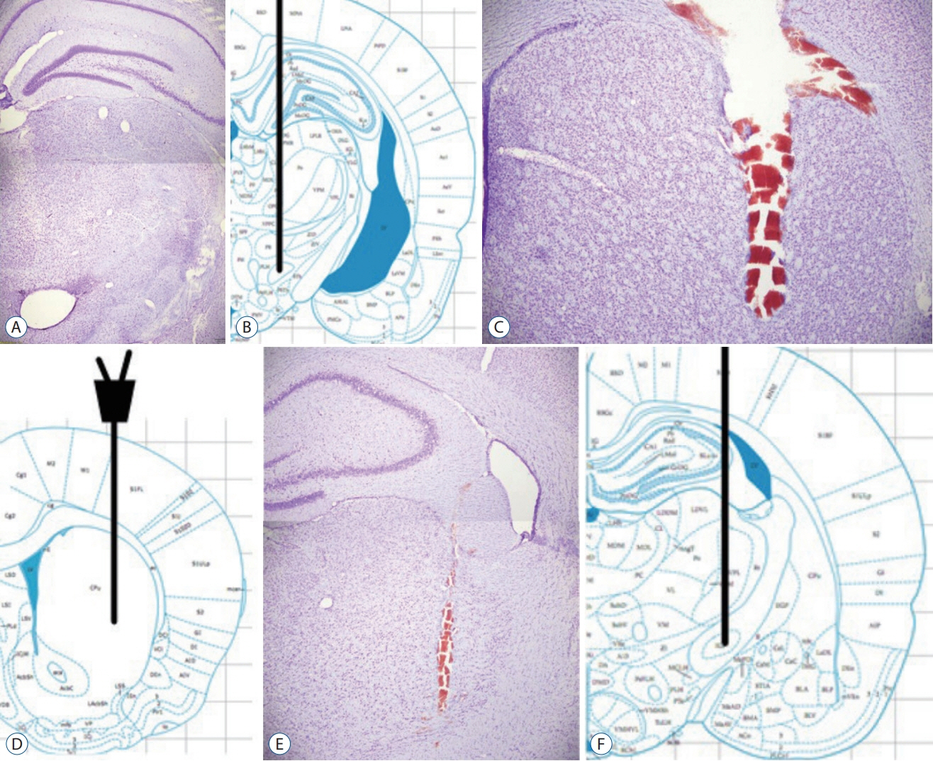
Fig. 5. Histological verification. The stereotaxic atlas from Paxinos [23] shows the location of MFB (B), dorsal striatum (D) and EPN (F). Representation of coronal sections verifies injection site of 6-hydroxydopamine in the MFB (A), position of the probe in the striatum (C) and the electrode in the EPN (E). Scale bars=200 μm. MFB : medial forebrain bundle, EPN : entopeduncular nucleus.
Reference
-
References
1. Abosch A, Kapur S, Lang AE, Hussey D, Sime E, Miyasaki H, et al. Stimulation of the subthalamic nucleus in Parkinson’s disease does not produce striatal dopamine release. Neurosurgery. 53:1095–1102. discussion 1102-1105. 2003.
Article2. Agnesi F, Blaha CD, Lin J, Lee KH. Local glutamate release in the rat ventral lateral thalamus evoked by high-frequency stimulation. J Neural Eng. 7:26009. 2010.
Article3. Bar-Gad I, Elias S, Vaadia E, Bergman H. Complex locking rather than complete cessation of neuronal activity in the globus pallidus of a 1-methyl-4-phenyl-1,2,3,6-tetrahydropyridine-treated primate in response to pallidal microstimulation. J Neurosci. 24:7410–7419. 2004.
Article4. Barnéoud P, Parmentier S, Mazadier M, Miquet JM, Boireau A, Dubédat P, et al. Effects of complete and partial lesions of the dopaminergic mesotelencephalic system on skilled forelimb use in the rat. Neuroscience. 67:837–848. 1995.
Article5. Boulet S, Lacombe E, Carcenac C, Feuerstein C, Sgambato-Faure V, Poupard A, et al. Subthalamic stimulation-induced forelimb dyskinesias are linked to an increase in glutamate levels in the substantia nigra pars reticulata. J Neurosci. 26:10768–10776. 2006.
Article6. Bruet N, Windels F, Carcenac C, Feuerstein C, Bertrand A, Poupard A, et al. Neurochemical mechanisms induced by high frequency stimulation of the subthalamic nucleus: increase of extracellular striatal glutamate and GABA in normal and hemiparkinsonian rats. J Neuropathol Exp Neurol. 62:1228–1240. 2003.
Article7. DeLong MR. Primate models of movement disorders of basal ganglia origin. Trends Neurosci. 13:281–285. 1990.
Article8. DeLong MR, Wichmann T. Basal ganglia circuits as targets for neuromodulation in Parkinson disease. JAMA Neurol. 72:1354–1360. 2015.
Article9. Gubellini P, Eusebio A, Oueslati A, Melon C, Kerkerian-Le Goff L, Salin P. Chronic high-frequency stimulation of the subthalamic nucleus and L-DOPA treatment in experimental parkinsonism: effects on motor behaviour and striatal glutamate transmission. Eur J Neurosci. 24:1802–1814. 2006.
Article10. Hazrati LN, Parent A. Differential patterns of arborization of striatal and subthalamic fibers in the two pallidal segments in primates. Brain Res. 598:311–315. 1992.
Article11. Jonkers N, Sarre S, Ebinger G, Michotte Y. MK801 suppresses the LDOPA-induced increase of glutamate in striatum of hemi-Parkinson rats. Brain Res. 926:149–155. 2002.
Article12. Lanciego JL, Gonzalo N, Castle M, Sanchez-Escobar C, Aymerich MS, Obeso JA. Thalamic innervation of striatal and subthalamic neurons projecting to the rat entopeduncular nucleus. Eur J Neurosci. 19:1267–1277. 2004.
Article13. Lang AE, Widner H. Deep brain stimulation for Parkinson’s disease: patient selection and evaluation. Mov Disord 17 Suppl. 3:S94–S101. 2002.
Article14. Lee KH, Kristic K, van Hoff R, Hitti FL, Blaha C, Harris B, et al. Highfrequency stimulation of the subthalamic nucleus increases glutamate in the subthalamic nucleus of rats as demonstrated by in vivo enzymelinked glutamate sensor. Brain Res. 1162:121–129. 2007.
Article15. Lee KJ, Shim I, Sung JH, Hong JT, Kim IS, Cho CB. Striatal glutamate and GABA after high frequency subthalamic stimulation in parkinsonian rat. J Korean Neurosurg Soc. 60:138–145. 2017.
Article16. Liu Y, Li W, Tan C, Liu X, Wang X, Gui Y, et al. Meta-analysis comparing deep brain stimulation of the globus pallidus and subthalamic nucleus to treat advanced Parkinson disease. J Neurosurg. 121:709–718. 2014.
Article17. McConnell GC, So RQ, Hilliard JD, Lopomo P, Grill WM. Effective deep brain stimulation suppresses low-frequency network oscillations in the basal ganglia by regularizing neural firing patterns. J Neurosci. 32:15657–15668. 2012.
Article18. McIntyre CC, Savasta M, Kerkerian-Le Goff L, Vitek JL. Uncovering the mechanism(s) of action of deep brain stimulation: activation, inhibition, or both. Clin Neurophysiol. 115:1239–1248. 2004.
Article19. Meissner W, Paul G, Reum T, Reese R, Sohr R, Morgenstern R, et al. The influence of pallidal deep brain stimulation on striatal dopaminergic metabolism in the rat. Neurosci Lett. 296:149–152. 2000.
Article20. Moon HC, Won SY, Kim EG, Kim HK, Cho CB, Park YS. Effect of optogenetic modulation on entopeduncular input affects thalamic discharge and behavior in an AAV2-α-synuclein-induced hemiparkinson rat model. Neurosci Lett. 662:129–135. 2018.
Article21. Odekerken VJ, van Laar T, Staal MJ, Mosch A, Hoffmann CF, Nijssen PC, et al. Subthalamic nucleus versus globus pallidus bilateral deep brain stimulation for advanced Parkinson’s disease (NSTAPS study): a randomised controlled trial. Lancet Neurol. 12:37–44. 2013.
Article22. Papa SM, Engber TM, Kask AM, Chase TN. Motor fluctuations in levodopa treated parkinsonian rats: relation to lesion extent and treatment duration. Brain Res. 662:69–74. 1994.
Article23. Paxinos G. The rat nervous system. ed 3. San Diego: Elsevier Academic Press;2004. p. 49–56.24. Sgambato-Faure V, Cenci MA. Glutamatergic mechanisms in the dyskinesias induced by pharmacological dopamine replacement and deep brain stimulation for the treatment of Parkinson’s disease. Prog Neurobiol. 96:69–86. 2012.
Article25. Shipton EA. Movement disorders and neuromodulation. Neurol Res Int. 2012:309431. 2012.
Article26. Stefani A, Fedele E, Galati S, Pepicelli O, Frasca S, Pierantozzi M, et al. Subthalamic stimulation activates internal pallidus: evidence from cGMP microdialysis in PD patients. Ann Neurol. 57:448–452. 2005.
Article27. Tan ZG, Zhou Q, Huang T, Jiang Y. Efficacies of globus pallidus stimulation and subthalamic nucleus stimulation for advanced Parkinson’s disease: a meta-analysis of randomized controlled trials. Clin Interv Aging. 11:777–786. 2016.28. Temel Y, Visser-Vandewalle V, Aendekerk B, Rutten B, Tan S, Scholtissen B, et al. Acute and separate modulation of motor and cognitive performance in parkinsonian rats by bilateral stimulation of the subthalamic nucleus. Exp Neurol. 193:43–52. 2005.
Article29. Wichmann T, Delong MR. Deep-brain stimulation for basal ganglia disorders. Basal Ganglia. 1:65–77. 2011.
Article
- Full Text Links
-
- Actions
-
Cited
- CITED
-
- Close
- Share
-
- Similar articles
-
- Striatal Glutamate and GABA after High Frequency Subthalamic Stimulation in Parkinsonian Rat
- Deep Brain Stimulation for the Treatment of Medically Intractable Epilepsy: a Review on Clinical Application
- The Changes of c-fos and c-jun after Capsaicine Treatment in the Rat Brain
- Deep Brain Stimulation of the Subthalamic and Pedunculopontine Nucleus in a Patient with Parkinson's Disease
- Deep Brain Stimulation for the Treatment of Movement Disorders
