Evaluation of factors influencing the success rate of orthodontic microimplants using panoramic radiographs
- Affiliations
-
- 1Postgraduate Orthodontic Program, Arizona School of Dentistry & Oral Health, A. T. Still University, Mesa, AZ, USA. jongmoon@wonkwang.ac.kr
- 2Graduate School of Dentistry, Kyung Hee University, Seoul, Korea, Korea.
- 3Department of Orthodontics and Wonkwang Dental Research Institute, University of Wonkwang School of Dentistry, Iksan, Korea.
- 4Department of Interdisciplinary Health Sciences, A. T. Still University, Mesa, AZ, USA.
- 5Department of Orthodontics, University of Wonkwang School of Dentistry, Iksan, Korea.
- 6Department of Orthodontics, Wonkwang University Sanbon Dental Hospital, Gunpo, Korea.
- KMID: 2441439
- DOI: http://doi.org/10.4041/kjod.2018.48.1.30
Abstract
OBJECTIVE
The purpose of this study was to investigate factors influencing the success rate of orthodontic microimplants (OMIs) using panoramic radiographs (PRs).
METHODS
We examined 160 OMIs inserted bilaterally in the maxillary buccal alveolar bone between the second premolars and first molars of 80 patients (51 women, 29 men; mean age, 18.0 ± 6.1 years) undergoing treatment for malocclusion. The angulation and position of OMIs, as well as other parameters, were measured on PRs. The correlation between each measurement and the OMI success rate was then evaluated.
RESULTS
The overall success rate was 85.0% (136/160). Age was found to be a significant predictor of implant success (p < 0.05), while sex, side of placement, extraction, and position of the OMI tip were not significant predictors (p > 0.05). The highest success rate was observed for OMIs with tips positioned on the interradicular midline (IRML; central position). Univariate analyses revealed that the OMI success rate significantly increased with an increase in the OMI length and placement height of OMI (p = 0.001). However, in simultaneous analyses, only length remained significant (p = 0.027). Root proximity, distance between the OMI tip and IRML, interradicular distance, alveolar crest width, distance between the OMI head and IRML, and placement angle were not factors for success. Correlations between the placement angle and all other measurements except root proximity were statistically significant (p < 0.05).
CONCLUSIONS
Our findings suggest that OMIs positioned more apically with a lesser angulation, as observed on PRs, exhibit high success rates.
Figure
-
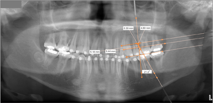
Figure 1 Panoramic radiograph incorporated in the V-Ceph imaging software (Osstem, Seoul, Korea).
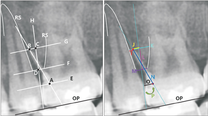
Figure 2 Reference lines and points (left) and linear and angular measurements (right) for an orthodontic microimplant (OMI) inserted in the maxillary buccal alveolar bone between the second premolar and first molar. OP, occlusal plane; A, head of OMI (HOMI); B, tip of OMI (TOMI); C, midpoint of the interradicular distance at the level of TOMI on a line parallel to OP; D, midpoint of the alveolar crest width; E, F, G, lines parallel to OP at the levels of HOMI (E), alveolar crest (F), and TOMI (G); H, interradicular midline drawn through C and D; RS, root surface; I, root proximity (RS to TOMI); J, distance between TOMI and the interradicular midline on a line parallel to OP; K, interradicular distance at the level of TOMI; L, placement height of OMI (HTOMI) measured from the midpoint of the interradicular distance to the midpoint of the alveolar crest width; M, alveolar crest width; N, length of OMI (LOMI) from TOMI to HOMI; O, distance between the interradicular midline and HOMI; P, placement angle measured between the long axis of OMI and the interradicular midline.
Reference
-
1. Min KI, Kim SC, Kang KH, Cho JH, Lee EH, Chang NY, et al. Root proximity and cortical bone thickness effects on the success rate of orthodontic micro-implants using cone beam computed tomography. Angle Orthod. 2012; 82:1014–1021.
Article2. Jung YR, Kim SC, Kang KH, Cho JH, Lee EH, Chang NY, et al. Placement angle effects on the success rate of orthodontic microimplants and other factors with cone-beam computed tomography. Am J Orthod Dentofacial Orthop. 2013; 143:173–181.
Article3. Lee MY, Park JH, Kim SC, Kang KH, Cho JH, Cho JW, et al. Bone density effects on the success rate of orthodontic microimplants evaluated with cone-beam computed tomography. Am J Orthod Dentofacial Orthop. 2016; 149:217–224.
Article4. Deguchi T, Nasu M, Murakami K, Yabuuchi T, Kamioka H, Takano-Yamamoto T. Quantitative evaluation of cortical bone thickness with computed tomographic scanning for orthodontic implants. Am J Orthod Dentofacial Orthop. 2006; 129:721.e7–721.e12.
Article5. Lim HJ, Eun CS, Cho JH, Lee KH, Hwang HS. Factors associated with initial stability of miniscrews for orthodontic treatment. Am J Orthod Dentofacial Orthop. 2009; 136:236–242.
Article6. Joo E. Radiographic evaluation of interdental distance for orthodontic miniscrew application [thesis]. Seoul: Yonsei University;2004.7. Kim SH, Yoon HG, Choi YS, Hwang EH, Kook YA, Nelson G. Evaluation of interdental space of the maxillary posterior area for orthodontic mini-implants with cone-beam computed tomography. Am J Orthod Dentofacial Orthop. 2009; 135:635–641.
Article8. Lim JE, Lim WH, Chun YS. Cortical bone thickness and root proximity at mandibular interradicular sites: implications for orthodontic mini-implant placement. Korean J Orthod. 2008; 38:397–406.
Article9. Kuroda S, Yamada K, Deguchi T, Hashimoto T, Kyung HM, Takano-Yamamoto T. Root proximity is a major factor for screw failure in orthodontic anchorage. Am J Orthod Dentofacial Orthop. 2007; 131:4 Suppl. S68–S73.
Article10. Schnelle MA, Beck FM, Jaynes RM, Huja SS. A radiographic evaluation of the availability of bone for placement of miniscrews. Angle Orthod. 2004; 74:832–837.11. Dudic A, Giannopoulou C, Leuzinger M, Kiliaridis S. Detection of apical root resorption after orthodontic treatment by using panoramic radiography and cone-beam computed tomography of super-high resolution. Am J Orthod Dentofacial Orthop. 2009; 135:434–437.
Article12. Cha JY, Mah J, Sinclair P. Incidental findings in the maxillofacial area with 3-dimensional cone-beam imaging. Am J Orthod Dentofacial Orthop. 2007; 132:7–14.
Article13. Nakagawa Y, Kobayashi K, Ishii H, Mishima A, Ishii H, Asada K, et al. Preoperative application of limited cone beam computerized tomography as an assessment tool before minor oral surgery. Int J Oral Maxillofac Surg. 2002; 31:322–326.
Article14. Bennemann R, Baxmann M, Keilig L, Reimann S, Braumann B, Bourauel C. Evaluating miniscrew position using orthopantomograms compared to cone-beam computed tomography. J Orofac Orthop. 2012; 73:236–248.
Article15. Larheim TA, Svanaes DB. Reproducibility of rotational panoramic radiography: mandibular linear dimensions and angles. Am J Orthod Dentofacial Orthop. 1986; 90:45–51.
Article16. Stewart JA, Heo G, Glover KE, Williamson PC, Lam EW, Major PW. Factors that relate to treatment duration for patients with palatally impacted maxillary canines. Am J Orthod Dentofacial Orthop. 2001; 119:216–225.
Article17. Yitschaky M, Haviv Y, Aframian DJ, Abed Y, Redlich M. Prediction of premolar tooth lengths based on their panoramic radiographic lengths. Dentomaxillofac Radiol. 2004; 33:370–372.
Article18. Hyomoto M, Kawakami M, Inoue M, Kirita T. Clinical conditions for eruption of maxillary canines and mandibular premolars associated with dentigerous cysts. Am J Orthod Dentofacial Orthop. 2003; 124:515–520.
Article19. Mckee IW, Glover KE, Williamson PC, Lam EW, Heo G, Major PW. The effect of vertical and horizontal head positioning in panoramic radiography on mesiodistal tooth angulations. Angle Orthod. 2001; 71:442–451.20. Kambylafkas P, Murdock E, Gilda E, Tallents RH, Kyrkanides S. Validity of panoramic radiographs for measuring mandibular asymmetry. Angle Orthod. 2006; 76:388–393.21. Mckee IW, Williamson PC, Lam EW, Heo G, Glover KE, Major PW. The accuracy of 4 panoramic units in the projection of mesiodistal tooth angulations. Am J Orthod Dentofacial Orthop. 2002; 121:166–175. quiz 192.
Article22. Stramotas S, Geenty JP, Petocz P, Darendeliler MA. Accuracy of linear and angular measurements on panoramic radiographs taken at various positions in vitro. Eur J Orthod. 2002; 24:43–52.
Article23. Nikneshan S, Sharafi M, Emadi N. Evaluation of the accuracy of linear and angular measurements on panoramic radiographs taken at different positions. Imaging Sci Dent. 2013; 43:191–196.
Article24. Choi BR, Choi DH, Huh KH, Yi WJ, Heo MS, Choi SC, et al. Clinical image quality evaluation for panoramic radiography in Korean dental clinics. Imaging Sci Dent. 2012; 42:183–190.
Article25. Alrbata RH, Momani MQ, Al-Tarawneh AM, Ihyasat A. Optimal force magnitude loaded to orthodontic microimplants: a finite element analysis. Angle Orthod. 2016; 86:221–226.
Article26. Papageorgiou SN, Zogakis IP, Papadopoulos MA. Failure rates and associated risk factors of orthodontic miniscrew implants: a meta-analysis. Am J Orthod Dentofacial Orthop. 2012; 142:577–595.e7.
Article27. Dalessandri D, Salgarello S, Dalessandri M, Lazzaroni E, Piancino M, Paganelli C, et al. Determinants for success rates of temporary anchorage devices in orthodontics: a meta-analysis (n > 50). Eur J Orthod. 2014; 36:303–313.
Article28. Mayoral G. Treatment results with light wires studied by panoramic radiography. Am J Orthod. 1982; 81:489–497.
Article29. Lucchesi MV, Wood RE, Nortjé CJ. Suitability of the panoramic radiograph for assessment of mesiodistal angulation of teeth in the buccal segments of the mandible. Am J Orthod Dentofacial Orthop. 1988; 94:303–310.
Article30. Ursi WJ, Almeida RR, Tavano O, Henriques JF. Assessment of mesiodistal axial inclination through panoramic radiography. J Clin Orthod. 1990; 24:166–173.31. Hong SB, Kusnoto B, Kim EJ, BeGole EA, Hwang HS, Lim HJ. Prognostic factors associated with the success rates of posterior orthodontic miniscrew implants: a subgroup meta-analysis. Korean J Orthod. 2016; 46:111–126.
Article32. Larheim TA, Eggen S. Determination of tooth length with a standardized paralleling technique and calibrated radiographic measuring film. Oral Surg Oral Med Oral Pathol. 1979; 48:374–378.
Article
- Full Text Links
-
- Actions
-
Cited
- CITED
-
- Close
- Share
-
- Similar articles
-
- Influence of late removal after treatment on the removal torque of microimplants
- External root resorption after orthodontic treatment: a study of contributing factors
- Optimization of orthodontic microimplant thread design
- Evaluation of Impacted Maxillary Canine Position Using Panoramic Radiographs and Cone-beam Computed Tomography
- Prediction of osteoporosis using fractal analysis on periapical and panoramic radiographs
