Global Synchronization Index as an Indicator for Tracking Cognitive Function Changes in a Traumatic Brain Injury Patient: A Case Report
- Affiliations
-
- 1Department of Physical Medicine and Rehabilitation, Hallym University Sacred Heart Hospital, Hallym University College of Medicine, Anyang, Korea. ohnsh@hallym.ac.kr
- KMID: 2440958
- DOI: http://doi.org/10.5535/arm.2019.43.1.106
Abstract
- Traumatic brain injury is a main cause of long-term neurological disability, and many patients suffer from cognitive impairment for a lengthy period. Cognitive impairment is a fatal malady to that limits active rehabilitation, and functional recovery in patients with traumatic brain injury. In severe cases, it is impossible to assess cognitive function precisely, and severe cognitive impairment makes it difficult to establish a rehabilitation plan, as well as evaluate the course of rehabilitation. Evaluation of cognitive function is essential for establishing a rehabilitation plan, as well as evaluating the course of rehabilitation. We report a case of the analysis of electroencephalography with global synchronization index and low-resolution brain electromagnetic tomography applied, for evaluation of cognitive function that was difficult with conventional tests, due to severe cognitive impairment in a 77-year-old male patient that experienced traumatic brain injury.
MeSH Terms
Figure
-
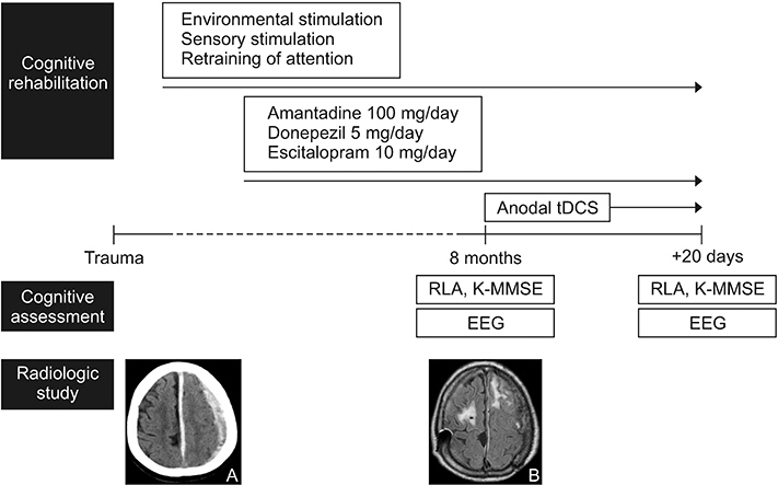
Fig. 1. Experimental design. Brain computed tomography taken at emergency room revealed subdural hemorrhage (A) and brain magnetic resonance imaging (MRI) taken at 8 months after trauma revealed prefrontal contusion (B). The patient received cognitive rehabilitation including cognitive therapy, medications, and transcranial direct current stimulation (tDCS). The cognitive function was assessed using Rancho Los Amigos (RLA) scale, Korean version of the Mini-Mental State Examination (K-MMSE), and electroencephalography (EEG) at the date that the brain MRI was taken and followed up after 20 days.
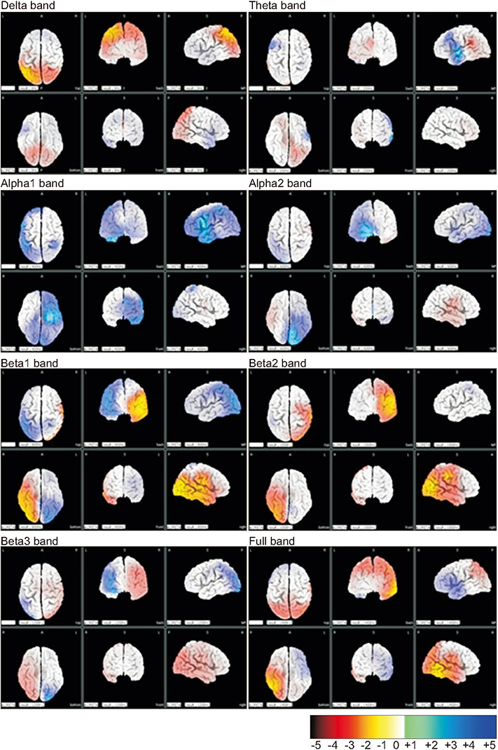
Fig. 2. LORETA analysis comparing the first and second EEG. Increased activities in the second EEG compared to the first EEG are highlighted in blue. Meanwhile, decreased activities between are highlighted in yellow-red. Each of the bands is composed of delta (1.5–4 Hz), theta (4–8 Hz), alpha1 (8–10 Hz), alpha2 (10–13 Hz), beta1 (13–18 Hz), beta2 (18–21 Hz), and beta3 (21–30 Hz) in ranges. LORETA, low resolution brain electromagnetic tomography; EEG, electroencephalography.
Reference
-
1. Li X, Cui D, Jiruska P, Fox JE, Yao X, Jefferys JG. Synchronization measurement of multiple neuronal populations. J Neurophysiol. 2007; 98:3341–8.
Article2. Cao C, Slobounov S. Alteration of cortical functional connectivity as a result of traumatic brain injury revealed by graph theory, ICA, and sLORETA analyses of EEG signals. IEEE Trans Neural Syst Rehabil Eng. 2010; 18:11–9.
Article3. Lee SH, Park YM, Kim DW, Im CH. Global synchronization index as a biological correlate of cognitive decline in Alzheimer’s disease. Neurosci Res. 2010; 66:333–9.
Article4. Liu T, Lin P, Chen Y, Wang J. Electroencephalogram synchronization analysis for attention deficit hyperactivity disorder children. Biomed Mater Eng. 2014; 24:1035–9.
Article5. Cui D, Liu J, Bian Z, Li Q, Wang L, Li X. Cortical source multivariate EEG synchronization analysis on amnestic mild cognitive impairment in type 2 diabetes. ScientificWorldJournal. 2014; 2014:523216.
Article6. Ianof JN, Fraga FJ, Ferreira LA, Ramos RT, Demario JL, Baratho R, et al. Comparative analysis of the electroencephalogram in patients with Alzheimer’s disease, diffuse axonal injury patients and healthy controls using LORETA analysis. Dement Neuropsychol. 2017; 11:176–85.
Article7. Klimesch W. EEG alpha and theta oscillations reflect cognitive and memory performance: a review and analysis. Brain Res Brain Res Rev. 1999; 29:169–95.
Article8. McCrea M, Prichep L, Powell MR, Chabot R, Barr WB. Acute effects and recovery after sport-related concussion: a neurocognitive and quantitative brain electrical activity study. J Head Trauma Rehabil. 2010; 25:283–92.9. Kang EK, Kim DY, Paik NJ. Transcranial direct current stimulation of the left prefrontal cortex improves attention in patients with traumatic brain injury: a pilot study. J Rehabil Med. 2012; 44:346–50.
Article10. Ulam F, Shelton C, Richards L, Davis L, Hunter B, Fregni F, et al. Cumulative effects of transcranial direct current stimulation on EEG oscillations and attention/working memory during subacute neurorehabilitation of traumatic brain injury. Clin Neurophysiol. 2015; 126:486–96.
Article
