Tenosynovial Bilateral Lipoma Arborescens of the Ankle in Adults
- Affiliations
-
- 1Department of Orthopedic Surgery, Inje University Busan Paik Hospital, Inje University College of Medicine, Busan, Korea. rmaqjawjr@gmail.com
- 2Department of Radiology, Inje University Busan Paik Hospital, Inje University College of Medicine, Busan, Korea.
- KMID: 2440583
- DOI: http://doi.org/10.14193/jkfas.2019.23.1.35
Abstract
- Lipoma arborescens or synovial lipomatosis is a rare disorder that is characterized by mature fat infiltration of the hypertrophic synovial villi, most frequently affecting the supra-patellar pouch of the knee. This paper presents a case of lipoma arborescens of the ankle joint bilaterally in an adult patient with involvement of both the intra-articular synovium and the synovial sheath of the tendons around the ankle.
MeSH Terms
Figure
-
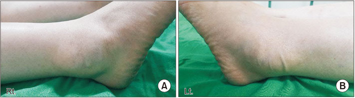
Figure 1 Photographs of the right ankle (A) and the left ankle (B) show soft-tissue swelling around lateral malleolus. Soft and movable mass-like lesion (A: 5×7 cm size, B: 4×6 cm size) was observed.
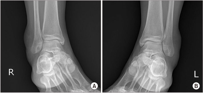
Figure 2 Internal oblique radiographics of the right ankle (A) and the left ankle (B) show soft tissue swelling of the postero-lateral ankle.
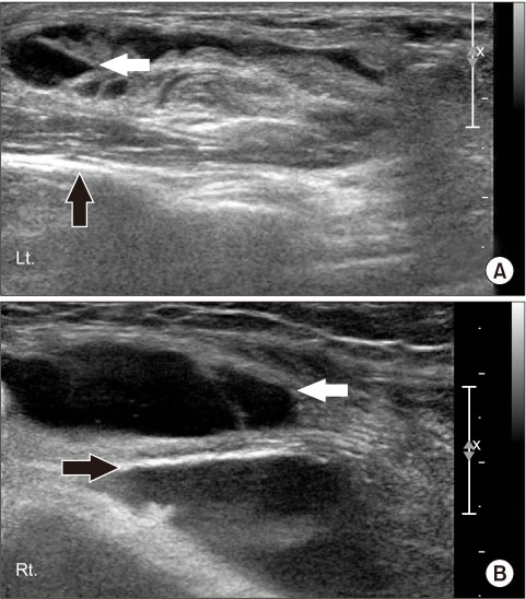
Figure 3 (A, B) Ultrasound scan, longitudinal view, shows encasement of the extensor tendons by the thickened synovium (black arrows). A Large amount of joint effusion in lateral aspect of both ankles (white arrows).
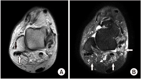
Figure 4 (A) Axial proton density-weighted magnetic resonance image showing high signal intensity within the peroneus brevis tendon at the postero-lateral malleolar level representing longitudinal tear (black arrow). The peroneus longus tendon (white arrow) is normal. (B) Axial T2-weighted fat-suppression magnetic resonance image at the corresponding level shows fluid collection with frond-like proliferations of the synovium of the involved tendon sheaths and ankle joint, with low signal intensity of the hyperplastic fat tissues (white arrows).
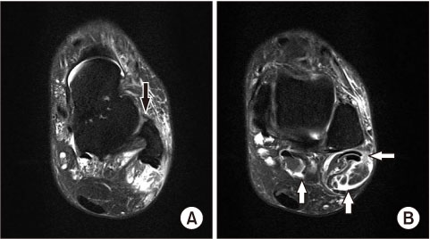
Figure 5 (A) The Anterior talofibular ligament is thin and has irregular margin and suspicious disruption of contour, meaning a chronic partial tear (black arrow). (B) Axial T2-weighted fat-suppression magnetic resonance image at the corresponding level shows fluid collection with frond-like proliferations of the synovium of the involved tendon sheaths and ankle joint, with low signal intensity of the hyperplastic fat tissues (white arrows).
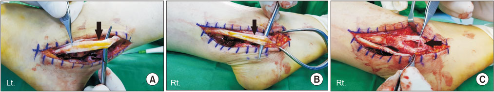
Figure 6 (A, B) Intraoperative photographs demonstrate abundant fat within the opened peroneal tendon sheath and partial tear of peroneus longus tendon in both ankles (black arrows). (C) There is a longitudinal split tear of the peroneus brevis tendon (black arrow).
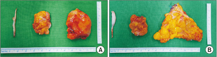
Figure 7 Gross photos of mass which are lipoma arborscence with abundant fat tissue and remnant of peroneus longus tendon in left ankle (A: 2×5 cm sized, 3.5 cm sized mass) and in right ankle (B: 2×2.5 cm sized, 6×6 cm sized mass).
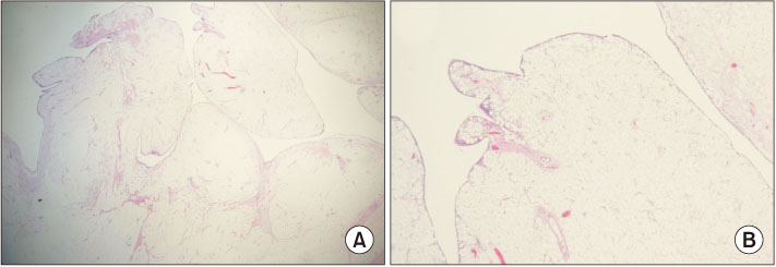
Figure 8 (A) Photomicrographs of excised synovium showing a lipomatous infiltrate within the synovium. On low power, a fatty infiltrate is seen in the synovium, giving it a lobular or villous contour (H&E stain, ×40). (B) High magnification shows mature adipose cells deep to a hyperplastic layer of synovial cells with scattered inflammatory cells and small plexiform vessels (H&E stain, ×40).
Cited by 1 articles
-
Intramuscular Giant Lipoma of the Anterior Compartment of the Ankle: A Case Report
Min Gu Jang, Jae Hwang Song, Jin Woong Yi, Dae Yeung Kim
J Korean Foot Ankle Soc. 2020;24(3):124-127. doi: 10.14193/jkfas.2020.24.3.124.
Reference
-
1. Vilanova JC, Barceló J, Villalón M, Aldomà J, Delgado E, Zapater I. MR imaging of lipoma arborescens and the associated lesions. Skeletal Radiol. 2003; 32:504–509.
Article2. Kakkar N, Vasishta RK, Anand H. Pathological case of the month. Synovial lipomatosis. Arch Pediatr Adolesc Med. 1999; 153:203–204.3. Laorr A, Peterfy CG, Tirman PF, Rabassa AE. Lipoma arborescens of the shoulder: magnetic resonance imaging findings. Can Assoc Radiol J. 1995; 46:311–313.4. Arzimanoglu A. Bilateral arborescent lipoma of the knee: a case report. J Bone Joint Surg Am. 1957; 39-A:976–979.5. Hallel T, Lew S, Bansal M. Villous lipomatous proliferation of the synovial membrane (lipoma arborescens). J Bone Joint Surg Am. 1988; 70:264–270.
Article6. Soler T, Rodríguez E, Bargiela A, Da Riba M. Lipoma arborescens of the knee: MR characteristics in 13 joints. J Comput Assist Tomogr. 1998; 22:605–609.
Article7. Siva C, Brasington R, Totty W, Sotelo A, Atkinson J. Synovial lipomatosis (lipoma arborescens) affecting multiple joints in a patient with congenital short bowel syndrome. J Rheumatol. 2002; 29:1088–1092.8. Huang GS, Lee HS, Hsu YC, Kao HW, Lee HH, Chen CY. Tenosynovial lipoma arborescens of the ankle in a child. Skeletal Radiol. 2006; 35:244–247.
Article9. Hughes M, Burtt S, Ramabhadran B. Lipoma arborescens-a rare cause of bilateral ankle tenosynovitis. Rheumatology (Oxford). 2018; 57:762–763.
Article
- Full Text Links
-
- Actions
-
Cited
- CITED
-
- Close
- Share
-
- Similar articles
-
- Lipoma Arborescens in the Hip Joint: A Case Report
- Lipoma Arborescens of Hip Joint: A Case Report
- Lipoma Arborescens in the Knee Joint: A Case Report
- Arthroscopic Treatment of the Lipoma Arborescens in All Compartments of the Knee: A Case Report
- The Arthroscopic Treatment of Lipoma Arborescens of Knee: A Case Report
