Recurred Plexiform Schwannoma of the Foot and Ankle
- Affiliations
-
- 1Department of Orthopaedic Surgery, Inje University Sanggye Paik Hospital, Seoul, Korea. orthoman@paik.ac.kr
- KMID: 2438972
- DOI: http://doi.org/10.4055/jkoa.2019.54.1.84
Abstract
- Schwannomas are benign neoplasms with a Schwann cell origin. A plexiform schwannoma is a rare variant of a schwannoma with a plexiform or multinodular growth pattern. The condition occurs mostly as a solitary lesion in the skin or subcutaneous tissue, or uncommonly located in the deep soft tissue. We report a rare case of recurred multiple plexiform schwannomas arising from the posterior tibial nerve and its branch, which was located in a deep anatomic location and accompanied by a bony deformity.
Keyword
MeSH Terms
Figure
-

Figure 1 Clinical photograph shows a previous curved surgical scar on the right ankle and foot. The mass is seen in the medial aspect of the right ankle, and atrophy of the plantar soft tissue is observed.
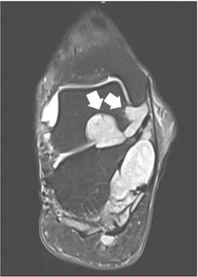
Figure 2 Coronal T2-weighted magnetic resonance imaging demonstrates space-occupying multiple lesions extending to the sinus tarsi with adjacent bony erosion (arrows).
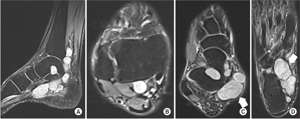
Figure 3 Sagittal T2-weighted (A) and axial T2-weighted magnetic resonance imagings at the level of the ankle (B), sinus tarsi (C), and plantar fascia (D) demonstrate multiple hyperintense lesions with bony deformity along the posterior tibial nerve, as well as the medial and lateral plantar nerve. The arrows indicate the Target sign.
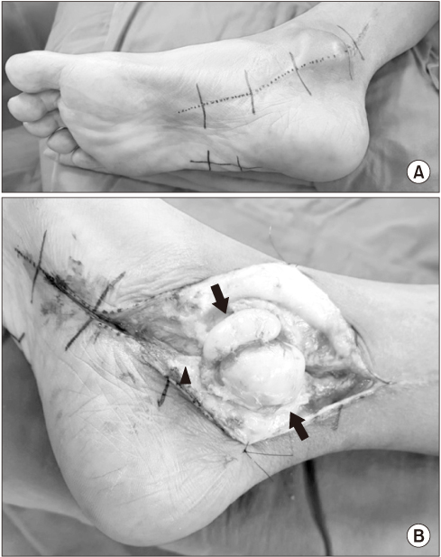
Figure 4 (A) Surgical incision marking. (B) Gross intraoperative appearance of two schwannomas (arrows) observed along the posterior tibial nerve (arrowheads).

Figure 5 Gross appearance of all distinct schwannomas in the right foot and ankle of the patient.
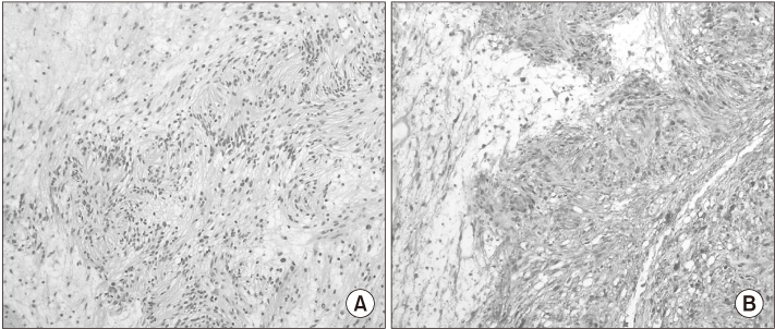
Figure 6 (A) Microscopic histological features show a characteristic biphasic pattern of Antoni A and B of neurogenic spindle cell proliferation (H&E, ×100). (B) Immunostaining for the S-100 protein is strong and diffuse (×10).
Reference
-
1. Ozdemir O, Ozsoy MH, Kurt C, Coskunol E, Calli I. Schwannomas of the hand and wrist: long-term results and review of the literature. J Orthop Surg (Hong Kong). 2005; 13:267–272.
Article2. Li XN, Cui JL, Christopasak SP, Kumar A, Peng ZG. Multiple plexiform schwannomas in the plantar aspect of the foot: case report and literature review. BMC Musculoskelet Disord. 2014; 15:342.
Article3. Hirose T, Scheithauer BW, Sano T. Giant plexiform schwannoma: a report of two cases with soft tissue and visceral involvement. Mod Pathol. 1997; 10:1075–1081.4. Agaram NP, Prakash S, Antonescu CR. Deep-seated plexiform schwannoma: a pathologic study of 16 cases and comparative analysis with the superficial variety. Am J Surg Pathol. 2005; 29:1042–1048.5. Mohammed SA, Pressman MM, Schmidt B, Babu N. Case presentations and review of plexiform schwannoma in the foot. J Foot Ankle Surg. 2014; 53:179–185.
Article6. Harkin JH, Arrington JH, Reed RJ. Benign plexiform schwannoma, a lesion distinct from plexiform neurofibroma. J Neuropathol Exp Neurol. 1978; 37:622.
Article7. Lee KH, Kim YS, Jeong CH, et al. Plexiform neurilemmoma unassociated with neurofibromatosis - 2 cases report - . J Korean Bone Joint Tumor Soc. 2005; 11:82–87.8. Jacobson JM, Felder JM 3rd, Pedroso F, Steinberg JS. Plexiform schwannoma of the foot: a review of the literature and case report. J Foot Ankle Surg. 2011; 50:68–73.
Article9. Nishio J, Mori S, Nabeshima K, Naito M. Successful enucleation of large multinodular/plexiform schwannoma of the foot and ankle. Springerplus. 2015; 17:260.
Article10. Yamada K, Harada M, Kunitoku N, Goto S, Kochi M, Ushio Y. MR imaging features of a scalp plexiform schwannoma. AJNR Am J Neuroradiol. 2004; 25:291–294.
