High-Resolution Magnetic Resonance Imaging Using Compressed Sensing for Intracranial and Extracranial Arteries: Comparison with Conventional Parallel Imaging
- Affiliations
-
- 1Department of Radiology and Research Institute of Radiology, University of Ulsan College of Medicine, Asan Medical Center, Seoul, Korea. dynamics79@gmail.com
- KMID: 2438279
- DOI: http://doi.org/10.3348/kjr.2018.0424
Abstract
OBJECTIVE
To compare conventional sensitivity encoding (SENSE) to compressed sensing plus SENSE (CS) for high-resolution magnetic resonance imaging (HR-MRI) of intracranial and extracranial arteries.
MATERIALS AND METHODS
HR-MRI was performed in 14 healthy volunteers. Three-dimensional T1-weighted imaging (T1WI) and proton density-weighted imaging (PD) were acquired using CS or SENSE under the same total acceleration factors (AF(t))-5.5, 6.8, and 9.7 for T1WI and 3.2, 4.0, and 5.8 for PD-to achieve reduced scanning times in comparison with the original imaging sequence (SENSE T1WI, AF(t) 3.5; SENSE PD, AF(t) 2.0) using the 3-tesla system. Two neuroradiologists measured signal-to-noise ratio (SNR) and contrast-to-noise ratio (CNR), and used visual scoring systems to assess image quality. Acceptable imaging was defined as a visual score ≥ 2. Repeated measures analysis of variance and Cochran's Q test were performed.
RESULTS
CS yielded better image quality and vessel delineation than SENSE in T1WI with AF(t) of 5.5, 6.8, and 9.7, and in PD with AF(t) of 5.8 (p < 0.05). CS T1WI with AF(t) of 5.5 and CS PD with AF(t) of 3.2 and 4.0 did not differ significantly from original imaging (p > 0.05). SNR and CNR in CS were higher than they were in SENSE, but lower than they were in the original images (p < 0.05). CS yielded higher proportions of acceptable imaging than SENSE (CS T1WI with AF(t) of 6.8 and PD with AF(t) of 5.8; p < 0.0167).
CONCLUSION
CS is superior to SENSE, and may be a reliable acceleration method for vessel HR-MRI using AF(t) of 5.5 for T1WI, and 3.2 and 4.0 for PD.
Keyword
MeSH Terms
Figure
-
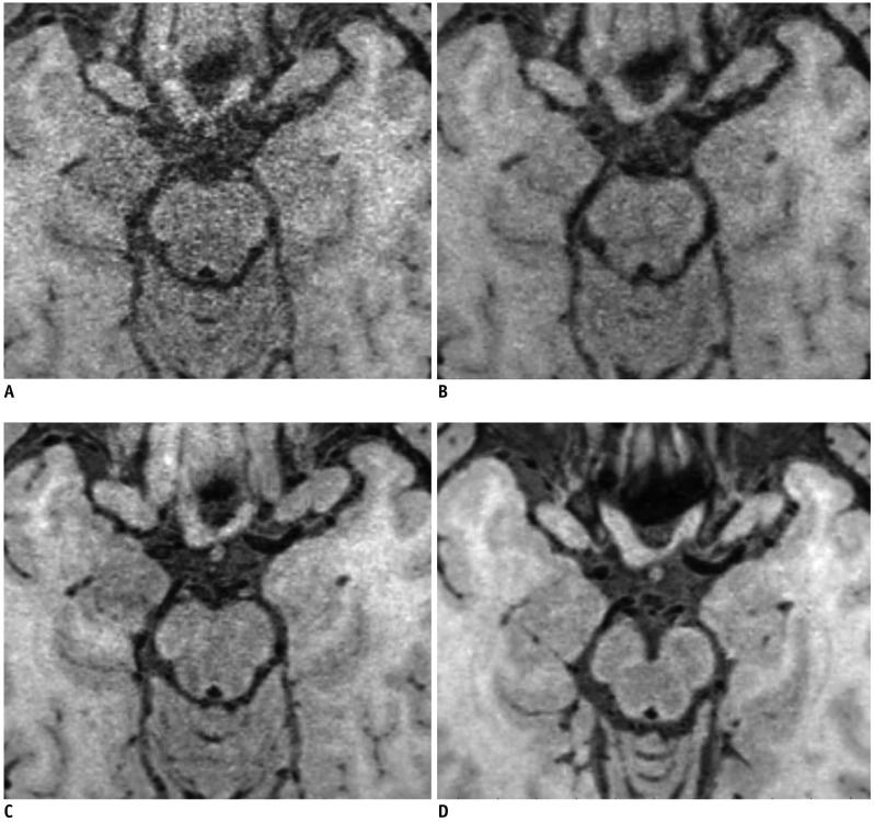
Fig. 1 Overall image quality and artifact were graded as follows.(A) Grade 0, poor image quality with large artifact, (B) grade 1, moderate image quality with moderate artifact, (C) grade 2, good image quality with slight artifact, and (D) grade 3, excellent image quality without artifact.
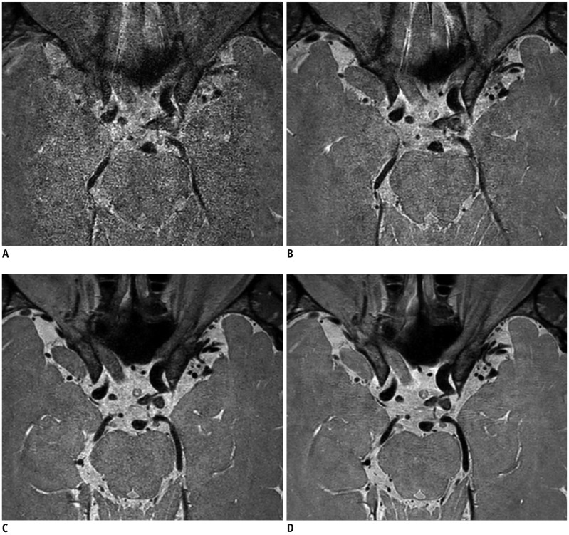
Fig. 2 Vessel delineations of outer contour and branching arteries were graded as follows.(A) Grade 0, less than 50% of vessel is visible, (B) grade 1, more than 50% of vessel is visible, (C) grade 2, vessel is delineated with adequate signal and contrast to lumen and CSF, and (D) grade 3, vessel is delineated with excellent signal and sharp contrast to lumen and CSF. CSF = cerebrospinal fluid
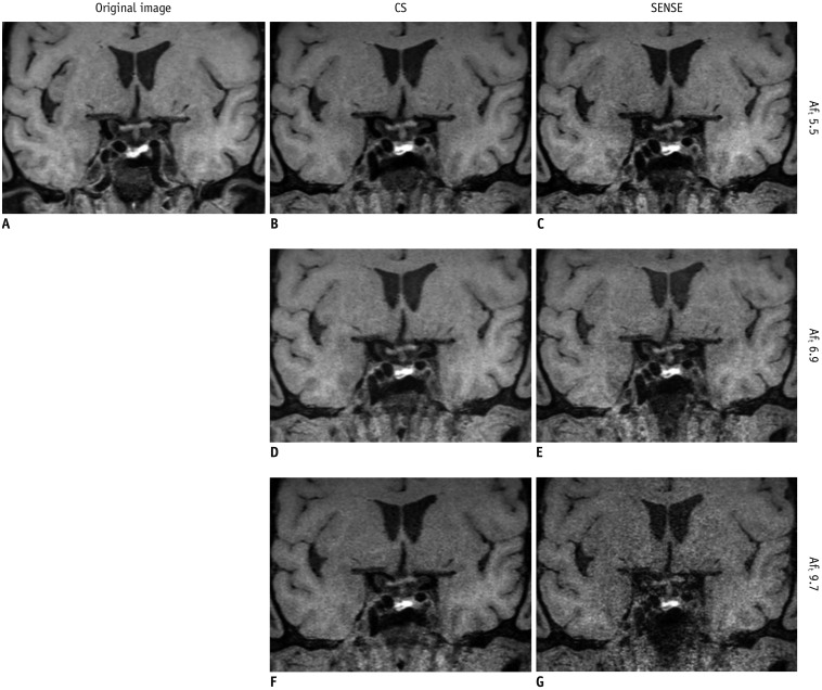
Fig. 3 Reconstructed coronal T1-weighted imaging of 59-year-old man.CS exhibits better image quality and vessel wall delineation than SENSE alone at each AFt. In comparison to original image (A), CS (B, D, F) exhibits better image quality and vessel wall delineation than SENSE alone (C, E, G) at each AFt. CS (B) and SENSE (C) images with AFt 5.5 may be clinically acceptable, whereas SENSE with AFt 9.7 (G) is uninterpretable. AFt = total acceleration factor, CS = compressed sensing plus SENSE, SENSE = sensitivity encoding

Fig. 4 Axial proton density-weighted imaging of 58-year-old man.CS yielded better image quality and vessel delineation for outer contour and branching arteries than SENSE alone at each AFt. In comparison to original image (A), CS (B, D, F) yielded better image quality and vessel delineation for outer contour and branching arteries than SENSE alone (C, E, G) at each AFt. CS (B, D) and SENSE (C, E) images with AFt 3.2 and 4.0 may be clinically acceptable, whereas SENSE with AFt 5.8 (G) is uninterpretable.
Reference
-
1. Mandell DM, Mossa-Basha M, Qiao Y, Hess CP, Hui F, Matouk C, Vessel Wall Imaging Study Group of the American Society of Neuroradiology, et al. Intracranial vessel wall MRI: principles and expert consensus recommendations of the American Society of Neuroradiology. AJNR Am J Neuroradiol. 2017; 38:218–222. PMID: 27469212.
Article2. Chung MS, Jung SC, Kim SO, Kim HS, Choi CG, Kim SJ, et al. Intracranial artery steno-occlusion: diagnosis by using two-dimensional spatially selective radiofrequency excitation pulse MR imaging. Radiology. 2017; 284:834–843. PMID: 28448235.
Article3. Lee NJ, Chung MS, Jung SC, Kim HS, Choi CG, Kim SJ, et al. Comparison of high-resolution MR imaging and digital subtraction angiography for the characterization and diagnosis of intracranial artery disease. AJNR Am J Neuroradiol. 2016; 37:2245–2250. PMID: 27659192.
Article4. Park JE, Jung SC, Lee SH, Jeon JY, Lee JY, Kim HS, et al. Comparison of 3D magnetic resonance imaging and digital subtraction angiography for intracranial artery stenosis. Eur Radiol. 2017; 27:4737–4746. PMID: 28500366.
Article5. Saam T, Raya JG, Cyran CC, Bochmann K, Meimarakis G, Dietrich O, et al. High resolution carotid black-blood 3T MR with parallel imaging and dedicated 4-channel surface coils. J Cardiovasc Magn Reson. 2009; 11:41. PMID: 19860875.
Article6. Itskovich VV, Mani V, Mizsei G, Aguinaldo JG, Samber DD, Macaluso F, et al. Parallel and nonparallel simultaneous multislice black-blood double inversion recovery techniques for vessel wall imaging. J Magn Reson Imaging. 2004; 19:459–467. PMID: 15065170.
Article7. Lustig M, Donoho D, Pauly JM. Sparse MRI: the application of compressed sensing for rapid MR imaging. Magn Reson Med. 2007; 58:1182–1195. PMID: 17969013.
Article8. Li B, Li H, Kong H, Dong L, Zhang J, Fang J. Compressed sensing based simultaneous black- and gray-blood carotid vessel wall MR imaging. Magn Reson Imaging. 2017; 38:214–223. PMID: 28109887.
Article9. Zhu C, Tian B, Chen L, Eisenmenger L, Raithel E, Forman C, et al. Accelerated whole brain intracranial vessel wall imaging using black blood fast spin echo with compressed sensing (CS-SPACE). MAGMA. 2018; 31:457–467. PMID: 29209856.
Article10. Fan Z, Yang Q, Deng Z, Li Y, Bi X, Song S, et al. Whole-brain intracranial vessel wall imaging at 3 Tesla using cerebrospinal fluid-attenuated T1-weighted 3D turbo spin echo. Magn Reson Med. 2017; 77:1142–1150. PMID: 26923198.
Article11. Ramesh N, Yoo JH, Sethi IK. Thresholding based on histogram approximation. IEE P-Vis Image Sign. 1995; 142:271–227.
Article12. Raju PDR, Neelima G. Image segmentation by using histogram thresholding. IJCSET. 2012; 2:776–779.13. Qiao Y, Steinman DA, Qin Q, Etesami M, Schär M, Astor BC, et al. Intracranial arterial wall imaging using three-dimensional high isotropic resolution black blood MRI at 3.0 Tesla. J Magn Reson Imaging. 2011; 34:22–23. PMID: 21698704.
Article14. Zhang Z, Fan Z, Carroll TJ, Chung Y, Weale P, Jerecic R, et al. Three-dimensional T2-weighted MRI of the human femoral arterial vessel wall at 3.0 Tesla. Invest Radiol. 2009; 44:619–626. PMID: 19692844.
Article15. Park JE, Han K, Sung YS, Chung MS, Koo HJ, Yoon HM, et al. Selection and reporting of statistical methods to assess reliability of a diagnostic test: conformity to recommended methods in a peer-reviewed journal. Korean J Radiol. 2017; 18:888–897. PMID: 29089821.
Article16. Kang HJ, Lee JM, Joo I, Hur BY, Jeon JH, Jang JY, et al. Assessment of malignant potential in intraductal papillary mucinous neoplasms of the pancreas: comparison between multidetector CT and MR imaging with MR cholangiopancreatography. Radiology. 2016; 279:128–139. PMID: 26517448.
Article17. Yang AC, Kretzler M, Sudarski S, Gulani V, Seiberlich N. Sparse reconstruction techniques in magnetic resonance imaging: methods, applications, and challenges to clinical adoption. Invest Radiol. 2016; 51:349–364. PMID: 27003227.
- Full Text Links
-
- Actions
-
Cited
- CITED
-
- Close
- Share
-
- Similar articles
-
- Uncover This Tech Term: Compressed Sensing Magnetic Resonance Imaging
- Advanced Methods in Dynamic Contrast Enhanced Arterial Phase Imaging of the Liver
- Intracranial Atherosclerosis: From Microscopy to High-Resolution Magnetic Resonance Imaging
- Vessel Wall Imaging of the Intracranial and Cervical Carotid Arteries
- Fast Imaging of Shoulder MR Arthrography With Compressed Sensing Accelerated Isovolumetric 3D-THRIVE Sequence: Comparing One Isovolumetric Scan With Multiplanar Reconstruction and Three Conventional MR Images
