A Case of Blastic Plasmacytoid Dendritic Cell Neoplasm
- Affiliations
-
- 1Department of Dermatology, Centro Hospitalar do Porto-Hospital de Santo Antonio, Porto, Portugal. teresap.almeida@hotmail.com
- 2Department of Hematology, Centro Hospitalar do Porto-Hospital de Santo Antonio, Porto, Portugal.
Abstract
- No abstract available.
MeSH Terms
Figure
-
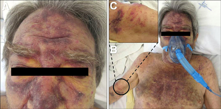
Fig. 1 (A, B) Features of the cutaneous lesions on the face and thorax. (C) Highlight of one firm subcutaneous nodule on the right arm.

Fig. 2 (A, B) Flow cytometry dot plots of peripheral blood (A) and skin (B) cells showing blastic plasmacytoid dendritic cells (blastic plasmacytoid dendritic cell neoplasm, red dots), respectively, expressing CD45 (dim), CD4 (dim), CD56, CD123, and HLA-DR. (C, D) Histopathological examination of a cutaneous lesion. (C) Diffuse infiltrate occupying the entire dermis and infiltrating the subcutaneous tissue (hematoxylin and eosin stain [H&E]; original magnification, ×4). (D) Higher amplification of the large blastic cells with large irregular nuclei (H&E; original magnification, ×40). SSC: side scatter; FSC: forward scatter.
Reference
-
1. Petrella T, Bagot M, Willemze R, Beylot-Barry M, Vergier B, Delaunay M, et al. Blastic NK-cell lymphomas (agranular CD4+CD56+ hematodermic neoplasms): a review. Am J Clin Pathol. 2005. 123:662–675.
Article2. Willemze R, Jaffe ES, Burg G, Cerroni L, Berti E, Swerdlow SH, et al. WHO-EORTC classification for cutaneous lymphomas. Blood. 2005. 105:3768–3785.
Article3. Faccheti F, Jones DM, Petrella T. Swerdlow SH, Campo E, Harris NL, Jaffe ES, Pileri SA, Stein H, editors. Blastic plasmacytoid dendritic cell neoplasm. WHO classification of tumours of haematopoietic and lymphoid tissues. 2008. 4th ed. Lyon: International Agency for Research on Cancer;145–147.4. Whittle AM, Howard MR. Skin lesions in plasmacytoid dendritic cell leukaemia. Br J Haematol. 2008. 140:121.
Article5. Magro CM, Porcu P, Schaefer J, Erter JW, Furman RR, Shitabata PK, et al. Cutaneous CD4+ CD56+ hematologic malignancies. J Am Acad Dermatol. 2010. 63:292–308.
Article6. Herling M, Jones D. CD4+/CD56+ hematodermic tumor: the features of an evolving entity and its relationship to dendritic cells. Am J Clin Pathol. 2007. 127:687–700.7. Garnache-Ottou F, Feuillard J, Ferrand C, Biichle S, Trimoreau F, Seilles E, et al. Extended diagnostic criteria for plasmacytoid dendritic cell leukaemia. Br J Haematol. 2009. 145:624–636.
Article8. Tsagarakis NJ, Kentrou NA, Papadimitriou KA, Pagoni M, Kokkini G, Papadaki H, et al. Acute lymphoplasmacytoid dendritic cell (DC2) leukemia: results from the Hellenic Dendritic Cell Leukemia Study Group. Leuk Res. 2010. 34:438–446.
Article9. Feuillard J, Jacob MC, Valensi F, Maynadié M, Gressin R, Chaperot L, et al. Clinical and biologic features of CD4(+) CD56(+) malignancies. Blood. 2002. 99:1556–1563.10. Reimer P, Rüdiger T, Kraemer D, Kunzmann V, Weissinger F, Zettl A, et al. What is CD4+CD56+ malignancy and how should it be treated? Bone Marrow Transplant. 2003. 32:637–646.
Article11. Jegalian AG, Buxbaum NP, Facchetti F, Raffeld M, Pittaluga S, Wayne AS, et al. Blastic plasmacytoid dendritic cell neoplasm in children: diagnostic features and clinical implications. Haematologica. 2010. 95:1873–1879.
Article
- Full Text Links
-
- Actions
-
Cited
- CITED
-
- Close
- Share
-
- Similar articles
-
- A Woman with Blastic Plasmacytoid Dendritic Cell Neoplasm
- Blastic Plasmacytoid Dendritic Cell Neoplasm Mimicking Traumatic Hematoma: A Case Report
- A Case of Blastic Plasmacytoid Dendritic Cell Neoplasm in Child
- A Case of Blastic Plasmacytoid Dendritic Cell Neoplasm with Mutations in DNMT3A, TET2, SRSF2, and ATRX Genes
- Plasmacytoid dendritic cell neoplasms
