J Cardiovasc Imaging.
2019 Jan;27(1):11-21. 10.4250/jcvi.2019.27.e3.
Gender Related Association between Arterial Stiffness and Aortic Root Geometry
- Affiliations
-
- 1Department of Internal Medicine, Boramae Medical Center, Seoul National University College of Medicine, Seoul, Korea. jooheezo@hanmail.net
- 2Department of Biostatistics, Boramae Medical Center, Seoul, Korea.
- KMID: 2433759
- DOI: http://doi.org/10.4250/jcvi.2019.27.e3
Abstract
- BACKGROUND
The gender-related change in aortic morphology by arterial stiffness has not been well studied. This study was performed to investigate the association between brachial-ankle pulse wave velocity (baPWV) and aortic root size according to gender.
METHODS
A total of 263 consecutive subjects (63.2 ± 10.6 years, 71.1% men) without overt cardiovascular disease who underwent both baPWV measurement and transthoracic echocardiography on the same day were retrospectively analyzed. The diameters of the aortic annulus (AN), sinus of Valsalva (SV), sinotubular junction (STJ), and ascending aorta (AA) were measured using 2-dimensional echocardiography.
RESULTS
The body surface area (BSA)-corrected diameters of AN, SV, STJ, and AA were significantly higher in women than in men. Univariable analyses showed that baPWV was significantly correlated with SV/BSA and STJ/BSA in men, and with SV/BSA, STJ/BSA, and AA/BSA in women (p < 0.05 for each). In men, however, these associations disappeared in multiple linear regression models after controlling for potential confounders (p > 0.05 for each). In women, the associations of baPWV with diameters of STJ/BSA (β = 0.407, p < 0.001) and AA/BSA (β = 0.391, p = 0.005) remained significant in the same multivariate models. Women-specific correlation between aortic root size and baPWV was also similarly demonstrated in age-matched analyses (n = 61 in each gender).
CONCLUSIONS
Among Korean adult without overt cardiovascular disease, the association between increased arterial stiffness and aortic root dilatation is stronger in women than in men.
MeSH Terms
Figure
-
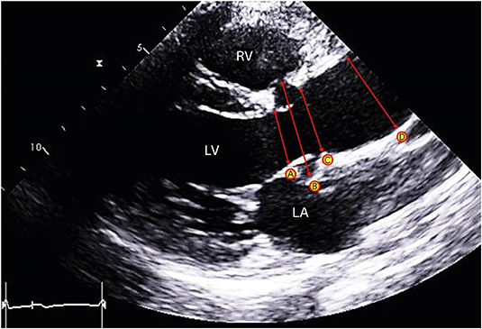
Figure 1 Measurements of aortic root size at 4 different levels using 2-dimensional transthoracic echocardiography. A indicates the annulus, B sinus of Valsalva, C the sinotubular junction, and D the proximal ascending aorta. LA: left atrium, LV: left ventricle, RV: right ventricle.
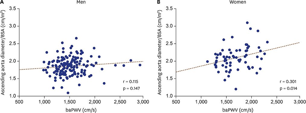
Figure 2 Scatter plots showing linear correlations between baPWV and BSA-corrected diameters of the ascending aorta. baPWV: brachial-ankle pulse wave velocity, BSA: body surface area.
Cited by 1 articles
-
Arterial Stiffness and Aortic Root Geometry: A Special Insight on Usual Materials
Byung Joo Sun
J Cardiovasc Imaging. 2019;27(1):22-23. doi: 10.4250/jcvi.2019.27.e11.
Reference
-
1. Lee HY, Oh BH. Aging and arterial stiffness. Circ J. 2010; 74:2257–2262.
Article2. Cavalcante JL, Lima JA, Redheuil A, Al-Mallah MH. Aortic stiffness: current understanding and future directions. J Am Coll Cardiol. 2011; 57:1511–1522.3. Vlachopoulos C, Aznaouridis K, Stefanadis C. Prediction of cardiovascular events and all-cause mortality with arterial stiffness: a systematic review and meta-analysis. J Am Coll Cardiol. 2010; 55:1318–1327.4. Yamashina A, Tomiyama H, Takeda K, et al. Validity, reproducibility, and clinical significance of noninvasive brachial-ankle pulse wave velocity measurement. Hypertens Res. 2002; 25:359–364.
Article5. Lee HS, Kim HL, Kim H, et al. Incremental prognostic value of brachial-ankle pulse wave velocity to single-photon emission computed tomography in patients with suspected coronary artery disease. J Atheroscler Thromb. 2015; 22:1040–1050.
Article6. Kim HL, Lee JM, Seo JB, et al. The effects of metabolic syndrome and its components on arterial stiffness in relation to gender. J Cardiol. 2015; 65:243–249.
Article7. Avolio A, Jones D, Tafazzoli-Shadpour M. Quantification of alterations in structure and function of elastin in the arterial media. Hypertension. 1998; 32:170–175.
Article8. Vriz O, Aboyans V, D'Andrea A, et al. Normal values of aortic root dimensions in healthy adults. Am J Cardiol. 2014; 114:921–927.
Article9. Nethononda RM, Lewandowski AJ, Stewart R, et al. Gender specific patterns of age-related decline in aortic stiffness: a cardiovascular magnetic resonance study including normal ranges. J Cardiovasc Magn Reson. 2015; 17:20.
Article10. Kim HL, Im MS, Seo JB, et al. The association between arterial stiffness and left ventricular filling pressure in an apparently healthy Korean population. Cardiovasc Ultrasound. 2013; 11:2.
Article11. Lancellotti P, Tribouilloy C, Hagendorff A, et al. European Association of Echocardiography recommendations for the assessment of valvular regurgitation. Part 1: aortic and pulmonary regurgitation (native valve disease). Eur J Echocardiogr. 2010; 11:223–244.
Article12. Kröner ES, Westenberg JJ, Kroft LJ, Brouwer NJ, van den Boogaard PJ, Scholte AJ. Coupling between MRI-assessed regional aortic pulse wave velocity and diameters in patients with thoracic aortic aneurysm: a feasibility study. Neth Heart J. 2015; 23:493–501.
Article13. Kröner ES, Scholte AJ, de Koning PJ, et al. MRI-assessed regional pulse wave velocity for predicting absence of regional aorta luminal growth in marfan syndrome. Int J Cardiol. 2013; 167:2977–2982.
Article14. Koullias G, Modak R, Tranquilli M, Korkolis DP, Barash P, Elefteriades JA. Mechanical deterioration underlies malignant behavior of aneurysmal human ascending aorta. J Thorac Cardiovasc Surg. 2005; 130:677–683.
Article15. Shim CY, Cho IJ, Yang WI, et al. Central aortic stiffness and its association with ascending aorta dilation in subjects with a bicuspid aortic valve. J Am Soc Echocardiogr. 2011; 24:847–852.
Article16. Seki M, Kurishima C, Kawasaki H, Masutani S, Senzaki H. Aortic stiffness and aortic dilation in infants and children with tetralogy of Fallot before corrective surgery: evidence for intrinsically abnormal aortic mechanical property. Eur J Cardiothorac Surg. 2012; 41:277–282.
Article17. Kojima T, Kuwata S, Kurishima C, et al. Aortic root dilatation and aortic stiffness in patients with single ventricular circulation. Circ J. 2014; 78:2507–2511.
Article18. Kosar F, Sincer I, Aksoy Y, Topal E, Cehreli S. Increased aortic stiffness in patients with coronary artery ectasia. Coron Artery Dis. 2005; 16:499–504.
Article19. Vriz O, Driussi C, Bettio M, Ferrara F, D'Andrea A, Bossone E. Aortic root dimensions and stiffness in healthy subjects. Am J Cardiol. 2013; 112:1224–1229.
Article20. Milan A, Tosello F, Naso D, et al. Ascending aortic dilatation, arterial stiffness and cardiac organ damage in essential hypertension. J Hypertens. 2013; 31:109–116.
Article21. Herrera VL, Decano JL, Giordano N, Moran AM, Ruiz-Opazo N. Aortic and carotid arterial stiffness and epigenetic regulator gene expression changes precede blood pressure rise in stroke-prone Dahl salt-sensitive hypertensive rats. PLoS One. 2014; 9:e107888.
Article22. Hickson SS, Butlin M, Graves M, et al. The relationship of age with regional aortic stiffness and diameter. JACC Cardiovasc Imaging. 2010; 3:1247–1255.
Article23. Vasan RS, Larson MG, Levy D. Determinants of echocardiographic aortic root size. The Framingham Heart Study. Circulation. 1995; 91:734–740.24. Coutinho T, Borlaug BA, Pellikka PA, Turner ST, Kullo IJ. Sex differences in arterial stiffness and ventricular-arterial interactions. J Am Coll Cardiol. 2013; 61:96–103.
Article25. Tanaka H, DeSouza CA, Seals DR. Absence of age-related increase in central arterial stiffness in physically active women. Arterioscler Thromb Vasc Biol. 1998; 18:127–132.
Article26. Vargas R, Wroblewska B, Rego A, Hatch J, Ramwell PW. Oestradiol inhibits smooth muscle cell proliferation of pig coronary artery. Br J Pharmacol. 1993; 109:612–617.
Article27. Ailawadi G, Eliason JL, Roelofs KJ, et al. Gender differences in experimental aortic aneurysm formation. Arterioscler Thromb Vasc Biol. 2004; 24:2116–2122.
Article28. Maurer G. Aortic regurgitation. Heart. 2006; 92:994–1000.
Article29. Weltert L, de Tullio MD, Afferrante L, et al. Annular dilatation and loss of sino-tubular junction in aneurysmatic aorta: implications on leaflet quality at the time of surgery. A finite element study. Interact Cardiovasc Thorac Surg. 2013; 17:8–12.
Article30. Grotenhuis HB, Ottenkamp J, Westenberg JJ, Bax JJ, Kroft LJ, de Roos A. Reduced aortic elasticity and dilatation are associated with aortic regurgitation and left ventricular hypertrophy in nonstenotic bicuspid aortic valve patients. J Am Coll Cardiol. 2007; 49:1660–1665.
Article
- Full Text Links
-
- Actions
-
Cited
- CITED
-
- Close
- Share
-
- Similar articles
-
- Arterial Stiffness and Aortic Root Geometry: A Special Insight on Usual Materials
- Validation of Three-Dimensional Echocardiography for Quantification of Aortic Root Geometry: Comparison with Multi-Detector Computed Tomography
- Arterial stiffness and hypertension
- Valve-Sparing Neo-Aortic Root Replacement for Neo-Aortic Root Dilatation 20 Years after Arterial Switch Operation for Transposition of the Great Arteries: A Case Report
- Aortic Valve Sparing Operations: A Review
