Reference Value for Infrapatellar Branch of Saphenous Nerve Conduction Study: Cadaveric and Clinical Study
- Affiliations
-
- 1Department of Rehabilitation Medicine, St. Paul's Hospital, College of Medicine, The Catholic University of Korea, Seoul, Korea. coltrane@catholic.ac.kr
- KMID: 2432203
- DOI: http://doi.org/10.5535/arm.2018.42.2.321
Abstract
OBJECTIVE
To determine the optimal stimulation and recording site for infrapatellar branch of saphenous nerve (IPBSN) conduction studies by a cadaveric study, and to confirm that obtained location is practically applicable to healthy adults.
METHODS
Twelve lower limbs from six cadavers were studied. We defined the optimal stimulation site as the point IPBSN exits the sartorius muscle and the distance or ratio were measured on the X- and Y-axis based on the line connecting the medial and lateral poles of the patella. We defined the optimal recording site as the point where the terminal branch met the line connecting inferior pole of patella and tibial tuberosity, and measured the distance from the inferior pole. Also, nerve conduction studies were performed with obtained location in healthy adults.
RESULTS
In optimal stimulation site, the mean value of X-coordinate was 55.50±6.10 mm, and the ratio of the Y-coordinate to the thigh length was 25.53%±5.40%. The optimal recording site was located 15.92±1.83 mm below the inferior pole of patella. In our sensory nerve conduction studies through this location, mean peak latency was 4.11±0.30 ms and mean amplitude was 4.16±1.49 µV.
CONCLUSION
The optimal stimulation site was located 5.0-6.0 cm medial to medial pole of the patella and 25% of thigh length proximal to the X-axis. The optimal recording site was located 1.5-2.0 cm below inferior pole of patella. We have also confirmed that this location is clinically applicable.
Keyword
MeSH Terms
Figure
-
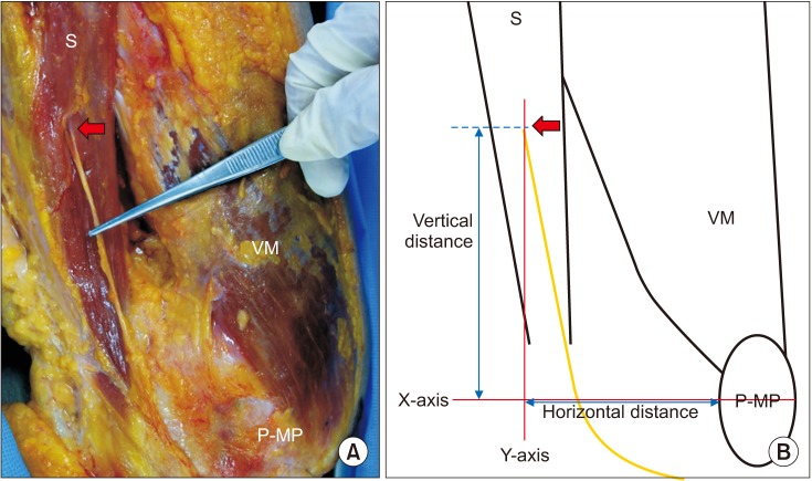
Fig. 1 (A) Anteromedial view of a dissected left knee showing the optimal stimulation site, where the IPBSN exit the sartorius muscle (arrow) in thigh. (B) Schematic illustration demonstrating anatomical landmarks and coordinates of stimulation site of IPBSN (arrow). The horizontal distance was from the medial pole of patella to the point where X- and Y-axis cross. The vertical distance was from the point where the nerve exiting the sartorius muscle to the point where X- and Y-axis cross. VM, vastus medialis muscle; S, sartorius muscle; P-MP, medial pole of patella; IPBSN, infrapatellar branch of saphenous nerve.
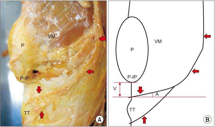
Fig. 2 (A) Anteromedial view of a dissected right knee showing the arc-like course and location of terminal branch of the IPBSN (arrow). (B) Schematic illustration demonstrating anatomical landmarks and relative location of the IPBSN (arrow). The vertical distance was from the inferior pole of the patella to the point where the terminal branch meets the reference line, connecting inferior pole of the patella and tibial tuberosity. The acute angle was the angle between terminal branch of the IPBSN and a perpendicular line to the reference line. VM, vastus medialis muscle; P, patella; P-IP, inferior pole of patella; TT, tibial tuberosity; V, vertical distance; AA, acute angle; IPBSN, infrapatellar branch of saphenous nerve.
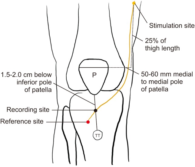
Fig. 3 Schematic illustration demonstrating nerve conduction study of right IPBSN. Stimulation was performed on the reference line on the X-axis at 50–60 mm medial to the medial pole of patella, and at 25% of thigh length on the Y-axis. Recording electrode was attached 1.5–2.0 cm below the inferior pole of the patella. Reference electrode was placed 4 cm distal to the recording electrode in consideration of the direction of the IPBSN. P, patella; TT, tibial tuberosity; IPBSN, infrapatellar branch of saphenous nerve.
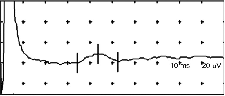
Fig. 4 Sensory nerve conduction study by obtained optimal stimulation and recording site for IPBSN (infrapatellar branch of saphenous nerve).
Reference
-
1. Kalthur SG, Sumalatha S, Nair N, Pandey AK, Sequeria S, Shobha L. Anatomic study of infrapatellar branch of saphenous nerve in male cadavers. Ir J Med Sci. 2015; 184:201–206. PMID: 24535194.
Article2. Le Corroller T, Lagier A, Pirro N, Champsaur P. Anatomical study of the infrapatellar branch of the saphenous nerve using ultrasonography. Muscle Nerve. 2011; 44:50–54. PMID: 21674521.
Article3. Bademkiran F, Obay B, Aydogdu I, Ertekin C. Sensory conduction study of the infrapatellar branch of the saphenous nerve. Muscle Nerve. 2007; 35:224–227. PMID: 17068766.
Article4. Ackmann T, Von During M, Teske W, Ackermann O, Muller P, Von Schulze Pellengahr C. Anatomy of the infrapatellar branch in relation to skin incisions and as the basis to treat neuropathic pain by cryodenervation. Pain Physician. 2014; 17:E339–E348. PMID: 24850115.5. Gali JC, Resina AF, Pedro G, Neto IA, Almagro MA, da Silva PA, et al. Importance of anatomically locating the infrapatellar branch of the saphenous nerve in reconstructing the anterior cruciate ligament using flexor tendons. Rev Bras Ortop. 2014; 49:625–629. PMID: 26229872.
Article6. Standring S. Gray's anatomy: the anatomical basis of clinical practice. 41st ed. New York: Elsevier;2016.7. Mistry D, O'Meeghan C. Fate of the infrapatellar branch of the saphenous nerve post total knee arthroplasty. ANZ J Surg. 2005; 75:822–824. PMID: 16174002.
Article8. Swanson AJ. The incidence of prepatellar neuropathy following medial meniscectomy. Clin Orthop Relat Res. 1983; 181:151–153.
Article9. Mochida H, Kikuchi S. Injury to infrapatellar branch of saphenous nerve in arthroscopic knee surgery. Clin Orthop Relat Res. 1995; 320:88–94.
Article10. Ikpeme JO, Gray C. Traumatic prepatellar neuralgia. Injury. 1995; 26:225–229. PMID: 7649620.
Article11. Saal JA, Dillingham MF, Gamburd RS, Fanton GS. The pseudoradicular syndrome. Lower extremity peripheral nerve entrapment masquerading as lumbar radiculopathy. Spine (Phila Pa 1976). 1988; 13:926–930. PMID: 2847334.
- Full Text Links
-
- Actions
-
Cited
- CITED
-
- Close
- Share
-
- Similar articles
-
- Conduction Studies of the Saphenous Nerve in Normal Subjects and Patients with Femoral Neuropathy
- Neurological Complication Following Total Knee Arthroplasty
- Morphology of saphenous nerve in cadavers: a guide to saphenous block and surgical interventions
- Preventing Lateral Skin Numbness after Medial Unicompartmental Knee Arthroplasty
- Pulsed Radiofrequency Neuromodulation for the Treatment of Saphenous Neuralgia
