Ann Rehabil Med.
2018 Apr;42(2):305-312. 10.5535/arm.2018.42.2.305.
Visual Evoked Potential in Children With Developmental Disorders: Correlation With Neurodevelopmental Outcomes
- Affiliations
-
- 1Department of Rehabilitation Medicine, Asan Medical Center, University of Ulsan College of Medicine, Seoul, Korea. iysung56@gmail.com
- KMID: 2432201
- DOI: http://doi.org/10.5535/arm.2018.42.2.305
Abstract
OBJECTIVE
To investigate the neurodevelopmental outcomes in children with developmental disorder according to visual evoked potential (VEP) results.
METHODS
We retrospectively analyzed children who visited our Department of Pediatric Rehabilitation Medicine with a chief complaint of developmental disability from January 2001 to July 2015. Of the 549 medical records reviewed, 322 children younger than 42 months who underwent both Bayley Scales of Infant and Toddler Development second edition (BSID-II) and VEP studies were enrolled. We compared the development of 182 children with normal VEP latency and 140 children with delayed VEP latency results using the BSID-II results. The Mann-Whitney U-test was used to analyze the differences between the two groups.
RESULTS
There were no significant differences in baseline characteristics between the two groups. The delayed VEP latency group showed a significant delay in BSID-II index scores and developmental quotients compared with the normal VEP latency group. In addition, a comparative analysis of developmental quotients of mental and psychomotor domains according to age (younger than 12 months, 12-23 months, and 24-42 months) revealed significantly lower values in children with delayed VEP latency compared to children with normal VEP latency, younger than 12 months and from 12 to 23 months.
CONCLUSION
Children with delayed VEP latency showed more developmental delay than children with normal VEP latency. It is suggested that VEP can be easily applied to children with suspected developmental delay when physicians have concerns about visual impairment. Furthermore, it is proposed that VEP results could provide an insight into children's development and serve as early indicators for consultation with an ophthalmologist for the existing problem.
MeSH Terms
Figure
-
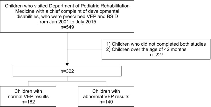
Fig. 1 Flowchart for the selection of study participants.

Fig. 2 Flash visual evoked potential record. (A) Normal latency. (B) Delayed latency.
Reference
-
1. Prechtl HF, Cioni G, Einspieler C, Bos AF, Ferrari F. Role of vision on early motor development: lessons from the blind. Dev Med Child Neurol. 2001; 43:198–201. PMID: 11263691.
Article2. Levtzion-Korach O, Tennenbaum A, Schnitzer R, Ornoy A. Early motor development of blind children. J Paediatr Child Health. 2000; 36:226–229. PMID: 10849221.
Article3. Vaizey MJ, Sanders MD, Wybar KC, Wilson J. Neurological abnormalities in congenital amaurosis of Leber. Review of 30 cases. Arch Dis Child. 1977; 52:399–402. PMID: 869570.
Article4. Cass HD, Sonksen PM, McConachie HR. Developmental setback in severe visual impairment. Arch Dis Child. 1994; 70:192–196. PMID: 7510945.
Article5. Odom JV, Bach M, Brigell M, Holder GE, McCulloch DL, Mizota A, et al. ISCEV standard for clinical visual evoked potentials: (2016 update). Doc Ophthalmol. 2016; 133:1–9.
Article6. Shepherd AJ, Saunders KJ, McCulloch DL, Dutton GN. Prognostic value of flash visual evoked potentials in preterm infants. Dev Med Child Neurol. 1999; 41:9–15. PMID: 10068044.
Article7. Vohr BR, Stephens BE, Higgins RD, Bann CM, Hintz SR, Das A, et al. Are outcomes of extremely preterm infants improving? Impact of Bayley assessment on outcomes. J Pediatr. 2012; 161:222–228. PMID: 22421261.
Article8. Lowe JR, Erickson SJ, Schrader R, Duncan AF. Comparison of the Bayley II Mental Developmental Index and the Bayley III Cognitive Scale: are we measuring the same thing? Acta Paediatr. 2012; 101:e55–e58. PMID: 22054168.
Article9. Moore T, Johnson S, Haider S, Hennessy E, Marlow N. Relationship between test scores using the second and third editions of the Bayley Scales in extremely preterm children. J Pediatr. 2012; 160:553–558. PMID: 22048046.
Article10. Niccols A, Latchman A. Stability of the Bayley mental scale of infant development with high risk infants. Br J Devel Disabil. 2002; 48:3–13.
Article11. Drislane FW. Visual evoked potentials. In : Blum AS, Rutkove SB, editors. The clinical neurophysiology primer. Totowa: Humana Press;2007. p. 461–474.12. Han SJ, Jeon SY. Comparison of visual evoked potentials between premature and full-term childrens. Acta Ophthalmol. 2012; 90(s249):F067.
Article13. Kato T, Watanabe K. Visual evoked potential in the newborn: does it have predictive value. Semin Fetal Neonatal Med. 2006; 11:459–463. PMID: 17070124.
Article14. Beverley DW, Smith IS, Beesley P, Jones J, Rhodes N. Relationship of cranial ultrasonography, visual and auditory evoked responses with neurodevelopmental outcome. Dev Med Child Neurol. 1990; 32:210–222. PMID: 2179002.
Article15. Ekert PG, Keenan NK, Whyte HE, Boulton J, Taylor MJ. Visual evoked potentials for prediction of neurodevelopmental outcome in preterm infants. Biol Neonate. 1997; 71:148–155. PMID: 9096893.
Article16. Pike AA, Marlow N. The role of cortical evoked responses in predicting neuromotor outcome in very preterm infants. Early Hum Dev. 2000; 57:123–135. PMID: 10735459.
Article17. Fulton AB, Hansen RM, Moskowitz A. Assessment of vision in infants and young children. In : Celesia GG, editor. Handbook of clinical neurophysiology : disorders of visual processing. Amsterdam: Elsevier;2005. p. 203–230.18. Harding GF. Flash visual evoked cortical potential in developmental delay. In : Heckenlively JR, Arden GB, editors. Principles and practice of clinical electrophysiology of vision. St. Louis: Mosby;1991. p. 585–588.
- Full Text Links
-
- Actions
-
Cited
- CITED
-
- Close
- Share
-
- Similar articles
-
- Comparative Analysis of Developmental Assessment, Evoked Potentials, Electroencephalography, and Brain MRI in Children with Cerebral Palsy
- Visual Evoked Potentials for Detecting Visual Pathway Abnormality
- Analysis of Brain Evoked Potential Study in Cerebral Palsy Patients
- Mental retardation and other neurodevelopmental disorders
- Abnormal Visual Evoked Potential Response from Hypoglycemic Encephalopathy in Two Neonates
