Ann Rehabil Med.
2018 Apr;42(2):286-295. 10.5535/arm.2018.42.2.286.
Relationship Between Functional Level and Muscle Thickness in Young Children With Cerebral Palsy
- Affiliations
-
- 1Department of Rehabilitation Medicine, Bundang Jesaeng General Hospital, Seongnam, Korea. drtaeim@gmail.com
- KMID: 2432199
- DOI: http://doi.org/10.5535/arm.2018.42.2.286
Abstract
OBJECTIVE
To investigate the relationship between functional level and muscle thickness (MT) of the rectus femoris (RF) and the gastrocnemius (GCM) in young children with cerebral palsy (CP).
METHODS
The study participants were comprised of 26 children (50 legs) with spastic CP, aged 3-6 years, and 25 age-matched children with typical development (TD, 50 legs). The MT of the RF, medial GCM, and lateral GCM was measured with ultrasound imaging. The functional level was evaluated using the Gross Motor Function Measurement-88 (GMFM-88), Gross Motor Function Classification System (GMFCS), and based on the mobility area of the Korean version of the Modified Barthel Index (K-MBI). The measurement of spasticity was evaluated with the Modified Ashworth Scale (MAS).
RESULTS
We note that the height, weight, body mass index, and MT of the RF, and the medial and lateral GCM were significantly higher in the TD group (p < 0.05). There was a direct relationship between MT of the RF and medial GCM and the GMFM-88, GMFCS, and mobility scores of the K-MBI in individuals with early CP. In addition, we have noted that there was a direct relationship between MT of the lateral GCM and the GMFM-88 and GMFCS. Although there was a tendency toward lower MT with increasing MAS ratings in the knee and ankle, the correlation was not statistically significant.
CONCLUSION
In young children with CP, MT of the RF and GCM was lower than in age-matched children with TD. Furthermore, it is noted with confidence that a significant positive correlation existed between MT and functional level as evaluated using the GMFM-88, GMFCS, and mobility area of K-MBI.
MeSH Terms
Figure
-
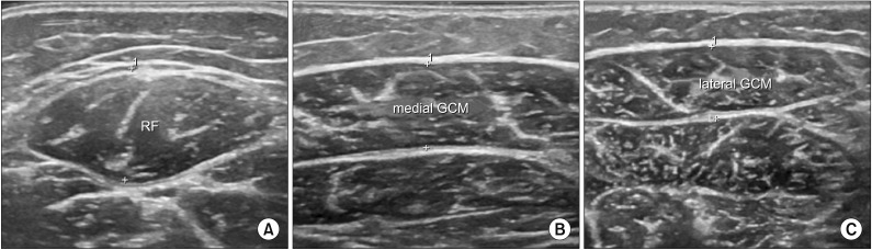
Fig. 1 Ultrasonographic measurement of muscle thickness: the longest distance between the upper muscular fascia and the lower muscular fascia. These figures show transverse images of (A) RF, (B) medial GCM, and (C) lateral GCM. RF, rectus femoris; GCM, gastrocnemius.
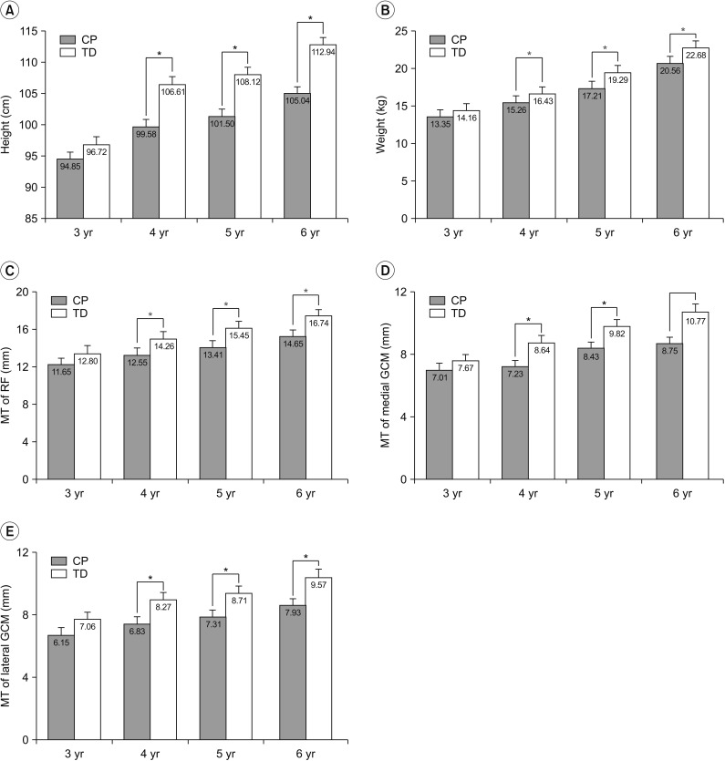
Fig. 2 Height, weight, and muscle thickness of the RF, medial GCM, and lateral GCM in cerebral palsy (CP) and typical development (TD) groups depending on age. (A) Height, (B) weight, (C) MT of RF, (D) MT of medial GCM, and (E) MT of lateral GCM. RF, rectus femoris; GCM, gastrocnemius; MT, muscle thickness. *p<0.05 by Mann-Whitney U-test independent t-test between CP and TD groups.
Reference
-
1. Bax M, Goldstein M, Rosenbaum P, Leviton A, Paneth N, Dan B, et al. Proposed definition and classification of cerebral palsy, April 2005. Dev Med Child Neurol. 2005; 47:571–576. PMID: 16108461.
Article2. Brown JK, Rodda J, Walsh EG, Wright GW. Neurophysiology of lower-limb function in hemiplegic children. Dev Med Child Neurol. 1991; 33:1037–1047. PMID: 1778340.
Article3. Elder GC, Kirk J, Stewart G, Cook K, Weir D, Marshall A, et al. Contributing factors to muscle weakness in children with cerebral palsy. Dev Med Child Neurol. 2003; 45:542–550. PMID: 12882533.
Article4. Poon DM, Hui-Chan CW. Hyperactive stretch reflexes, co-contraction, and muscle weakness in children with cerebral palsy. Dev Med Child Neurol. 2009; 51:128–135. PMID: 19018843.
Article5. Wiley ME, Damiano DL. Lower-extremity strength profiles in spastic cerebral palsy. Dev Med Child Neurol. 1998; 40:100–107. PMID: 9489498.
Article6. Riad J, Haglund-Akerlind Y, Miller F. Power generation in children with spastic hemiplegic cerebral palsy. Gait Posture. 2008; 27:641–647. PMID: 17951060.
Article7. Ross SA, Engsberg JR. Relationships between spasticity, strength, gait, and the GMFM-66 in persons with spastic diplegia cerebral palsy. Arch Phys Med Rehabil. 2007; 88:1114–1120. PMID: 17826455.
Article8. Shortland A. Muscle deficits in cerebral palsy and early loss of mobility: can we learn something from our elders. Dev Med Child Neurol. 2009; (51 Suppl 4):59–63. PMID: 19740211.
Article9. Moreau NG, Teefey SA, Damiano DL. In vivo muscle architecture and size of the rectus femoris and vastus lateralis in children and adolescents with cerebral palsy. Dev Med Child Neurol. 2009; 51:800–806. PMID: 19459913.
Article10. Barrett RS, Lichtwark GA. Gross muscle morphology and structure in spastic cerebral palsy: a systematic review. Dev Med Child Neurol. 2010; 52:794–804. PMID: 20477832.
Article11. Barber L, Hastings-Ison T, Baker R, Barrett R, Lichtwark G. Medial gastrocnemius muscle volume and fascicle length in children aged 2 to 5 years with cerebral palsy. Dev Med Child Neurol. 2011; 53:543–548. PMID: 21506995.12. Ohata K, Tsuboyama T, Haruta T, Ichihashi N, Kato T, Nakamura T. Relation between muscle thickness, spasticity, and activity limitations in children and adolescents with cerebral palsy. Dev Med Child Neurol. 2008; 50:152–156. PMID: 18201305.
Article13. Moreau NG, Simpson KN, Teefey SA, Damiano DL. Muscle architecture predicts maximum strength and is related to activity levels in cerebral palsy. Phys Ther. 2010; 90:1619–1630. PMID: 20847035.
Article14. Ohata K, Tsuboyama T, Ichihashi N, Minami S. Measurement of muscle thickness as quantitative muscle evaluation for adults with severe cerebral palsy. Phys Ther. 2006; 86:1231–1239. PMID: 16959671.
Article15. Bénard MR, Becher JG, Harlaar J, Huijing PA, Jaspers RT. Anatomical information is needed in ultrasound imaging of muscle to avoid potentially substantial errors in measurement of muscle geometry. Muscle Nerve. 2009; 39:652–665. PMID: 19291798.
Article16. Russell DJ, Avery LM, Rosenbaum PL, Raina PS, Walter SD, Palisano RJ. Improved scaling of the gross motor function measure for children with cerebral palsy: evidence of reliability and validity. Phys Ther. 2000; 80:873–885. PMID: 10960935.
Article17. Palisano R, Rosenbaum P, Walter S, Russell D, Wood E, Galuppi B. Development and reliability of a system to classify gross motor function in children with cerebral palsy. Dev Med Child Neurol. 1997; 39:214–223. PMID: 9183258.
Article18. Jung HY, Park BK, Shin HS, Kang YK, Pyun SB, Paik NJ, et al. Development of the Korean version of Modified Barthel Index (K-MBI): multi-center study for subjects with stroke. J Korean Acad Rehabil Med. 2007; 31:283–297.19. Shin WH, Kwon JY, Park HS, Jee SH, Jeong BL, Nam CM. Comparison of appropriateness of the Korean version of MBI and the Korea version of the PEDI for evaluation the activities of daily living on infants with cerebral palsy. J Korean Soc Occup Ther. 2013; 21:125–137.20. Malaiya R, McNee AE, Fry NR, Eve LC, Gough M, Shortland AP. The morphology of the medial gastrocnemius in typically developing children and children with spastic hemiplegic cerebral palsy. J Electromyogr Kinesiol. 2007; 17:657–663. PMID: 17459729.
Article21. Stackhouse SK, Binder-Macleod SA, Lee SC. Voluntary muscle activation, contractile properties, and fatigability in children with and without cerebral palsy. Muscle Nerve. 2005; 31:594–601. PMID: 15779003.
Article22. Mizner RL, Snyder-Mackler L. Altered loading during walking and sit-to-stand is affected by quadriceps weakness after total knee arthroplasty. J Orthop Res. 2005; 23:1083–1090. PMID: 16140191.
Article23. de Boer MD, Maganaris CN, Seynnes OR, Rennie MJ, Narici MV. Time course of muscular, neural and tendinous adaptations to 23 day unilateral lower-limb suspension in young men. J Physiol. 2007; 583(Pt 3):1079–1091. PMID: 17656438.
Article24. de Boer MD, Seynnes OR, di Prampero PE, Pisot R, Mekjavic IB, Biolo G, et al. Effect of 5 weeks horizontal bed rest on human muscle thickness and architecture of weight bearing and non-weight bearing muscles. Eur J Appl Physiol. 2008; 104:401–407. PMID: 18320207.25. Park ES, Sim E, Rha DW, Jung S. Estimation of gastrocnemius muscle volume using ultrasonography in children with spastic cerebral palsy. Yonsei Med J. 2014; 55:1115–1122. PMID: 24954345.
Article26. Miyatani M, Kanehisa H, Kuno S, Nishijima T, Fukunaga T. Validity of ultrasonograph muscle thickness measurements for estimating muscle volume of knee extensors in humans. Eur J Appl Physiol. 2002; 86:203–208. PMID: 11990727.
Article
- Full Text Links
-
- Actions
-
Cited
- CITED
-
- Close
- Share
-
- Similar articles
-
- Effect of Vibration Exercise Application on the Trunk Muscle Thickness in Children with Spastic Cerebral Palsy
- Leg Length Discrepancy in Children with Hemiplegic Cerebral Palsy
- Ultrasonographic Measurement of Gastrocnemius Muscle Thickness in Spastic Cerebral Palsy and Influencing Factors
- Nutritional Status of Children with Cerebral Palsy
- Analysis of Increased Myotatic Reflex in Children with Spastic Cerebral Palsy
