Complete mouth rehabilitation with vertical dimension increase in patient with extremely worn dentition
- Affiliations
-
- 1Department of Prosthodontic Dentistry, Veterans Health Service Medical Center, Seoul, Republic of Korea. ilovedua@naver.com
- KMID: 2432002
- DOI: http://doi.org/10.4047/jkap.2019.57.1.49
Abstract
- Generalized severely worn dentition causes occlusal disharmony, esthetic problems, and temporomandibular joint disorders. In order to solve these problems, it is necessary to make a precise analysis of vertical dimension and treatment plans considering it. This case report demonstrates the complete mouth rehabilitation of a 58-year-old male patient with a lot of worn teeth by increasing vertical dimension. Provisional restorations were cemented and after 4 months of evaluation for patient's compliance, permanent prostheses were fabricated. With these treatments, functionally and esthetically satisfactory results were obtained.
MeSH Terms
Figure
-

Fig. 1 Extraoral photograph before treatment. (A) Frontal view, (B) Frontal view smile, (C) Lateral view.
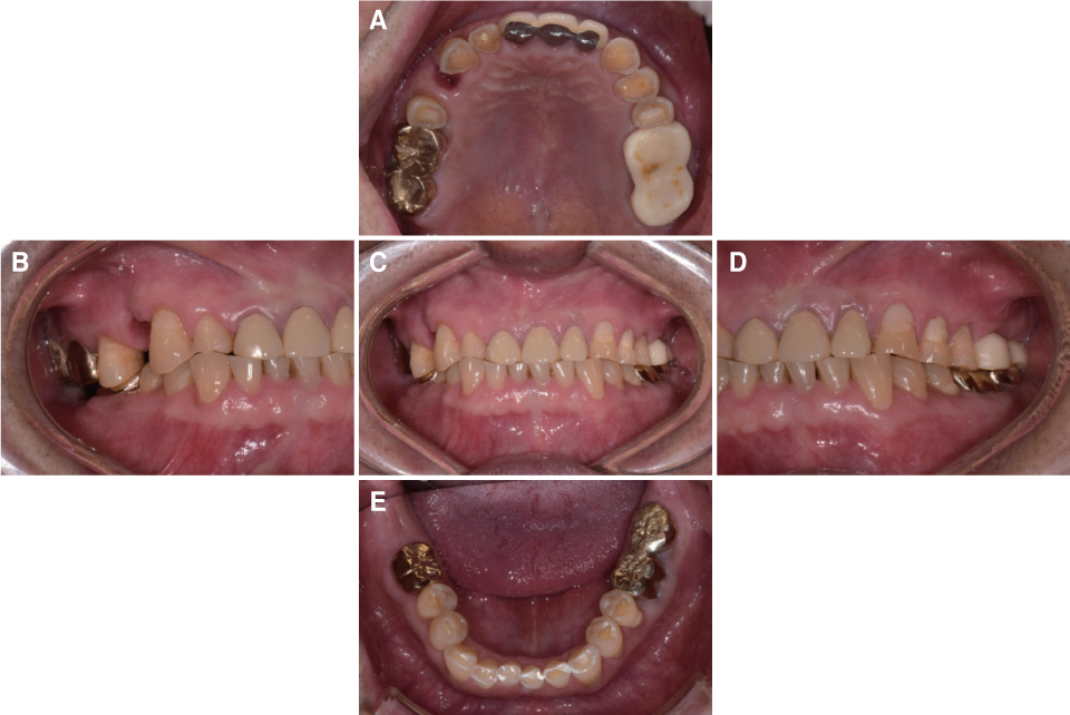
Fig. 2 Intraoral photograph before treatment. (A) Upper, (B) Right, (C) Frontal, (D) Left, (E) Lower.
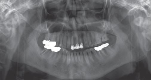
Fig. 3 Panoramic radiograph before treatment.

Fig. 4 TMJ series before treatment. (A) Rt. close, (B) Rt. opening, (C) Lt. opening, (D) Lt. close.
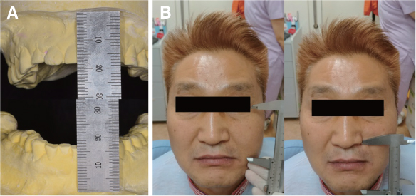
Fig. 5 Determining vertical dimension of occlusion. (A) Diagnostic cast, (B) Willis method.
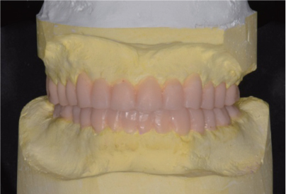
Fig. 6 Diagnostic wax-up with an increase of 3 mm of incisal guide pin.
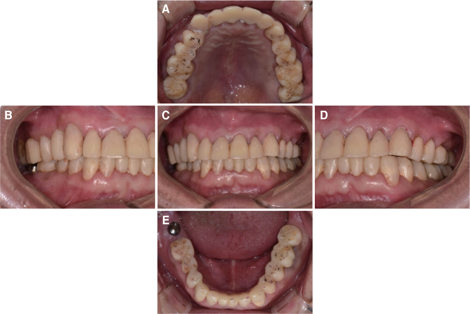
Fig. 7 Provisional restoration. (A) Upper, (B) Right, (C) Frontal, (D) Left, (E) Lower.
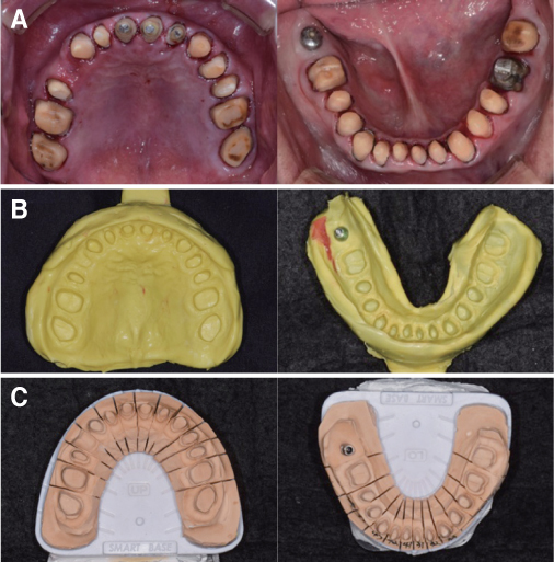
Fig. 8 (A) Tooth preparation, (B) Final impression taking, (C) Die preparation.

Fig. 9 (A) Full contour wax-up, (B) Cut back, (C) Metal coping fabrication.
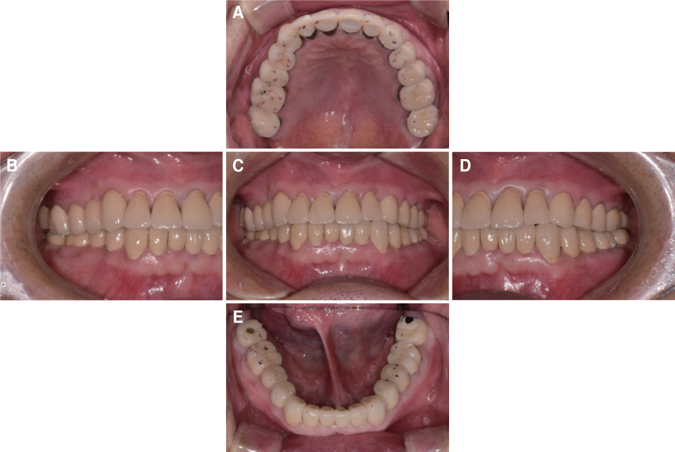
Fig. 10 Final prosthesis. (A) Upper, (B) Right, (C) Frontal, (D) Left, (E) Lower.
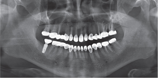
Fig. 11 Panoramic radiograph after treatment.
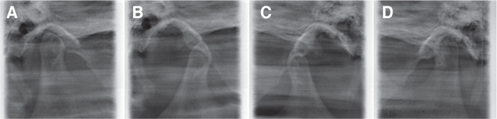
Fig. 12 TMJ series after treatment. (A) Rt. close, (B) Rt. opening, (C) Lt. opening, (D) Lt. close.

Fig. 13 Extraoral photograph after treatment. (A) Frontal view, (B) Frontal view smile, (C) Lateral view.
Reference
-
1. Murphy T. Compensatory mechanisms in facial height adjustment to functional tooth attrition. Aust Dent J. 1959; 4:312–323.
Article2. Rivera-Morales WC, Mohl ND. Relationship of occlusal vertical dimension to the health of the masticatory system. J Prosthet Dent. 1991; 65:547–553.
Article3. Briggs P, Bishop K. Fixed prostheses in the treatment of tooth wear. Eur J Prosthodont Restor Dent. 1997; 5:175–180.4. Hemmings KW, Darbar UR, Vaughan S. Tooth wear treated with direct composite restorations at an increased vertical dimension: results at 30 months. J Prosthet Dent. 2000; 83:287–293.
Article5. Sato S, Hotta TH, Pedrazzi V. Removable occlusal overlay splint in the management of tooth wear: a clinical report. J Prosthet Dent. 2000; 83:392–395.
Article6. Dahl BL, Krogstad O. The effect of a partial bite-raising splint on the inclination of upper and lower front teeth. Acta Odontol Scand. 1983; 41:311–314.
Article7. Ramfjord SP, Blankenship JR. Increased occlusal vertical dimension in adult monkeys. J Prosthet Dent. 1981; 45:74–83.
Article8. Dawson PE. Functional occlusion: From TMJ to smile design. St. Louis: Mosby Elsevier;2007. p. 430–452.9. Turner KA, Missirlian DM. Restoration of the extremely worn dentition. J Prosthet Dent. 1984; 52:467–474.
Article10. Willis FM. Features of the face involved in full denture prosthesis. Dent Cosmos. 1935; 77:851–854.11. Silverman MM. The speaking method in measuring vertical dimension. J Prosthet Dent. 2001; 85:427–431.
Article12. Park JH, Jeong CM, Jeon YC, Lim JS. A study on the occlusal plane and the vertical dimension in Korean adults with natural dentition. J Korean Acad Prosthodont. 2005; 43:41–51.13. Rivera-Morales WC, Mohl ND. Restoration of the vertical dimension of occlusion in the severely worn dentition. Dent Clin North Am. 1992; 36:651–664.14. Verrett RG. Analyzing the etiology of an extremely worn dentition. J Prosthodont. 2001; 10:224–233.
Article15. Jagger DC, Harrison A. An in vitro investigation into the wear effects of selected restorative materials on enamel. J Oral Rehabil. 1995; 22:275–281.
Article16. Lussi A. Dental erosion clinical diagnosis and case history taking. Eur J Oral Sci. 1996; 104:191–198.
Article17. Jacobi R, Shillingburg HT Jr, Duncanson MG Jr. A comparison of the abrasiveness of six ceramic surfaces and gold. J Prosthet Dent. 1991; 66:303–309.
Article18. Dahl BL, Carlsson GE, Ekfeldt A. Occlusal wear of teeth and restorative materials. A review of classification, etiology, mechanisms of wear, and some aspects of restorative procedures. Acta Odontol Scand. 1993; 51:299–311.
Article19. Abduo J, Lyons K. Clinical considerations for increasing occlusal vertical dimension: a review. Aust Dent J. 2012; 57:2–10.
Article
- Full Text Links
-
- Actions
-
Cited
- CITED
-
- Close
- Share
-
- Similar articles
-
- Full-mouth rehabilitation without changing the vertical dimension in patient with worn dentition
- Full mouth rehabilitation with extremely worn dentition
- Full mouth rehabilitation using zirconia crown in severe worn dentition: a case report
- Full mouth rehabilitation with vertical dimension increase in patient with loss of anterior guidance due to maxillary anterior teeth wear: A case report
- Mouth rehabilitation of a patient with severely worn dentition with vertical dimension increase
