A Case of Thoracic Extradural Chordoid Meningioma: Focusing on Radiologic Features
- Affiliations
-
- 1Department of Radiology, Dong-A University Medical Center, Busan, Korea. hdhdoc@naver.com
- 2Department of Pathology, Dong-A University Medical Center, Busan, Korea.
- 3Department of Neurology, Dong-A University Medical Center, Busan, Korea.
- KMID: 2431112
- DOI: http://doi.org/10.13104/imri.2018.22.4.260
Abstract
- Chordoid meningioma, an uncommon subtype of meningioma, occurs rarely in the spine. In this case report, the authors present a case of spinal chordoid meningioma in a young female patient, and include a detailed description of imaging findings and a literature review.
MeSH Terms
Figure
-
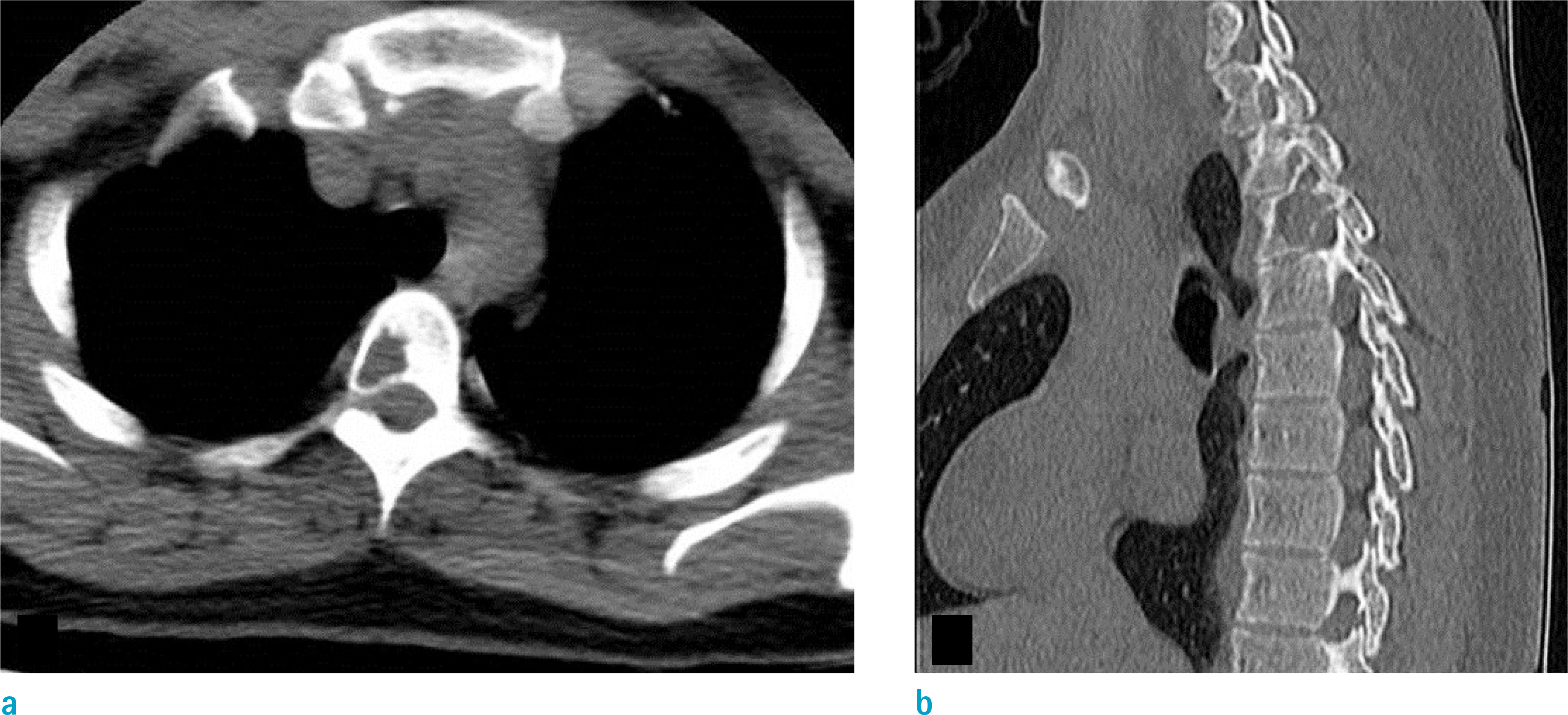
Fig. 1. Axial (a) and sagittal (b) CT scans show a well-defined osteolytic lesion without sclerotic margin, involving the T3 vertebral body, right pedicle and lamina.

Fig. 2. Sagittal fat suppressed T2-weighted image (a), sagittal T1-weighted image (b), sagittal gadolinium-enhanced T1-weighted image (c), sagittal diffusion-weighted image (d), sagittal apparent diffusion coefficient map (e), axial T2-weighted image (f), and axial gadolinium-enhanced T1-weighted image (g). There is an irregularly shaped mass with high signal intensity on T2-weighted images (a, f) and intermediate to low signal intensity on T1-weighted image (b). The mass is located in the extradural space of the T2–3 level, and invades the posterior components of the T3 vertebra. Due to the mass effect, the spinal cord is displaced to the left side. On gadolinium-enhanced T1-weighted image (c, g), the mass shows heterogeneous, but avid enhancement with fuzzy margins. The mass shows slight hyperintensity on diffusion-weighted image (d), and apparent diffusion coefficient map (e).

Fig. 3. Histopathologic findings. (a) Chords or trabeculae of eosinophilic epithelioid tumor cells in mucoid matrix (Hematoxylin & Eosin [H&E] stain, × 100). (b) Typical meningothelial meningioma-like focus (dotted circle, H&E stain, × 150). (c) Immunohistochemical staining for vimentin highlights the ribbon-like architecture (× 100). (d) Positivity for EMA is typically patchy in meningioma (× 100).
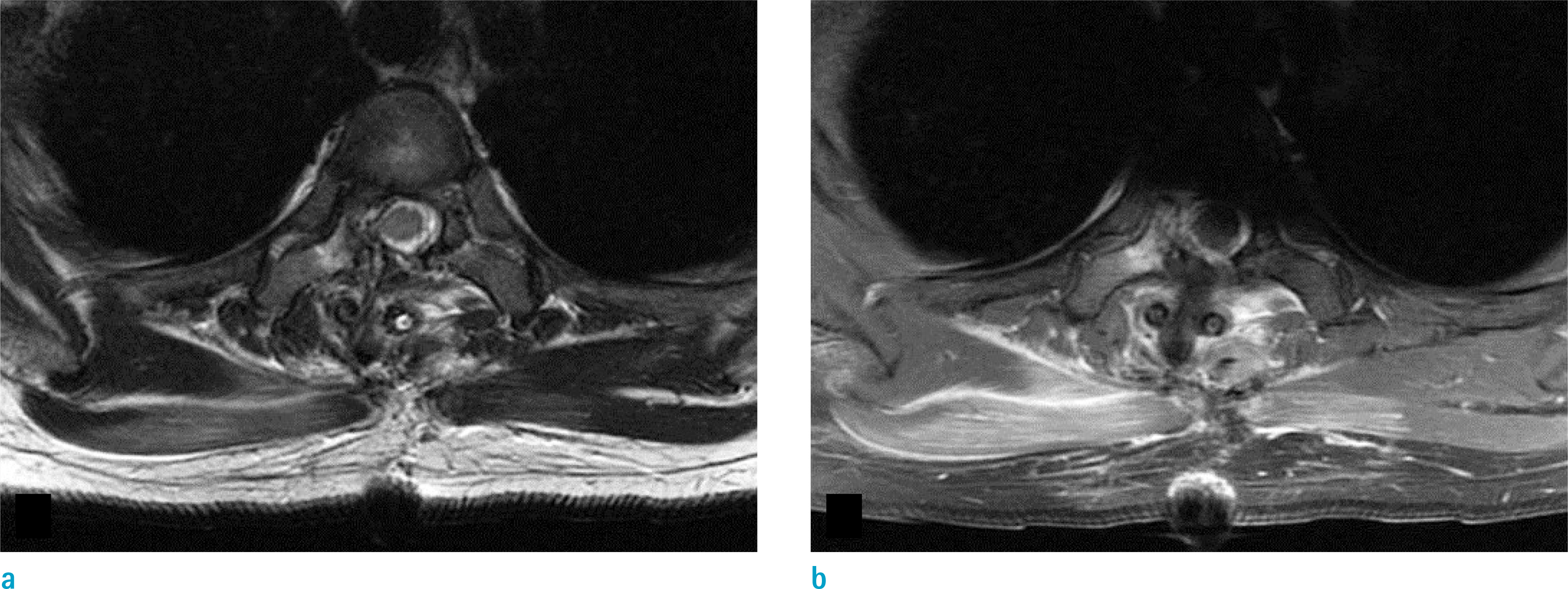
Fig. 4. On postoperative MRI, ill-defined hyperintense areas are noted in the T3 vertebra on axial T2-weighted image (a), and enhancing lesion in the same area on axial gadolinium-enhanced T1-weighted image (b), suggesting residual tumor.
Reference
-
References
1. Sadashiva N, Poyuran R, Mahadevan A, Bhat DI, Somanna S, Devi BI. Chordoid meningioma: a clinicopathological study of an uncommon variant of meningioma. J Neurooncol. 2018; 137:575–582.
Article2. Tulloch I, Georges H, Phadke R, Hardwidge C. A thoracic extradural chordoid meningioma: a unique case report and literature review. Br J Neurosurg. 2018. 1–3.
Article3. Yang Y, Li D, Cao XY, et al. Clinical features, treatment, and prognostic factors of chordoid meningioma: radiological and pathological features in 60 cases of chordoid meningioma. World Neurosurg. 2016; 93:198–207.
Article4. Wu L, Yang T, Fang J, Zhang J, Xu Y. Spinal chordoid meningioma in a child: a case report and review of the literature. Oncol Lett. 2015; 10:3727–3731.
Article5. Ibrahim A, Galloway M, Leung C, Revesz T, Crockard A. Cervical spine chordoid meningioma. Case report. J Neurosurg Spine. 2005; 2:195–198.6. Couce ME, Aker FV, Scheithauer BW. Chordoid meningioma: a clinicopathologic study of 42 cases. Am J Surg Pathol. 2000; 24:899–905.7. Badinand B, Morel C, Kopp N, Tran Min VA, Cotton F. Dumbbell-shaped epidural capillary hemangioma. AJNR Am J Neuroradiol. 2003; 24:190–192.8. Lee JW, Cho EY, Hong SH, et al. Spinal epidural hemangiomas: various types of MR imaging features with histopathologic correlation. AJNR Am J Neuroradiol. 2007; 28:1242–1248.
Article9. Dehcordi SR, Ricci A, Chiominto A, De Paulis D, Di Vitantonio H, Galzio RJ. Dorsal extradural meningioma: case report and literature review. Surg Neurol Int. 2016; 7:76.
Article10. Pond JB, Morgan TG, Hatanpaa KJ, Yetkin ZF, Mickey BE, Mendelsohn DB. Chordoid meningioma: differentiating a rare World Health Organization grade II tumor from other meningioma histologic subtypes using MRI. AJNR Am J Neuroradiol. 2015; 36:1253–1258.
Article
- Full Text Links
-
- Actions
-
Cited
- CITED
-
- Close
- Share
-
- Similar articles
-
- Primary Pulmonary Chordoid Meningioma
- An Extradural En-Plaque Meningioma Involving Thoracic Spine
- Extradural Spinal Lymphoplasmacyte-Rich Meningioma in the Thoracic Spine: A Case Report and Literature Review
- Spinal Extradural Meningioma: A Case Report and Review of the Literature
- Purely Extradural Spinal Meningioma of the Cervical Spine
