Korean J Orthod.
2019 Jan;49(1):41-48. 10.4041/kjod.2019.49.1.41.
A comparative study of the reproducibility of landmark identification on posteroanterior and anteroposterior cephalograms generated from cone-beam computed tomography scans
- Affiliations
-
- 1Department of Periodontology, School of Dentistry, Chonnam National University, Gwangju, Korea.
- 2Department of Orthodontics, School of Dentistry, Chonnam National University, Gwangju, Korea. ortholkm@jnu.ac.kr
- KMID: 2431079
- DOI: http://doi.org/10.4041/kjod.2019.49.1.41
Abstract
OBJECTIVE
This in-vivo study aimed to compare landmark identification errors in anteroposterior (AP) and posteroanterior (PA) cephalograms generated from cone-beam computed tomography (CBCT) scan data in order to examine the feasibility of using AP cephalograms in clinical settings.
METHODS
AP and PA cephalograms were generated from CBCT scans obtained from 25 adults. Four experienced and four inexperienced examiners were selected depending on their experience levels in analyzing frontal cephalograms. They identified six cephalometric landmarks on AP and PA cephalograms. The errors incurred in positioning the cephalometric landmarks on the AP and PA cephalograms were calculated by using the straight-line distance and the horizontal and vertical components as parameters.
RESULTS
Comparison of the landmark identification errors in CBCT-generated frontal cephalograms revealed that landmark-dependent differences were greater than experience- or projection-dependent differences. Comparisons of landmark identification errors in the horizontal and vertical directions revealed larger errors in identification of the crista galli and anterior nasal spine in the vertical direction and the menton in the horizontal direction, in comparison with the other landmarks. Comparison of landmark identification errors between the AP and PA projections in CBCT-generated images revealed a slightly higher error rate in the AP projections, with no inter-examiner differences. Statistical testing of the differences in landmark identification errors between AP and PA cephalograms showed no statistically significant differences for all landmarks.
CONCLUSIONS
The reproducibility of CBCT-generated AP cephalograms is comparable to that of PA cephalograms; therefore, AP cephalograms can be generated reliably from CBCT scan data in clinical settings.
Figure
-
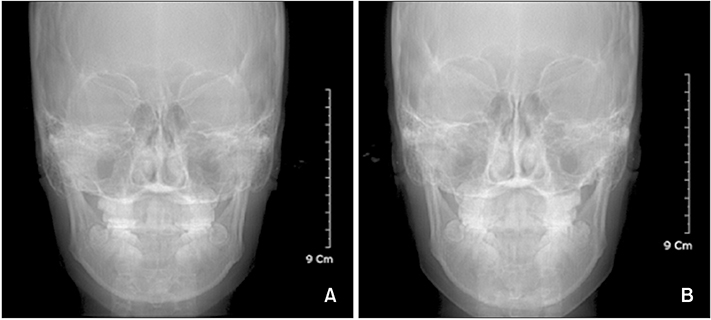
Figure 1 Frontal cephalograms generated from conebeam computed tomography scan data using the OnDemand3D program (Cybermed Inc., Seoul, Korea). A, Anteroposterior projection; B, postero-anterior projection.
Cited by 1 articles
-
Evaluation of a multi-stage convolutional neural network-based fully automated landmark identification system using cone-beam computed tomography-synthesized posteroanterior cephalometric images
Min-Jung Kim, Yi Liu, Song Hee Oh, Hyo-Won Ahn, Seong-Hun Kim, Gerald Nelson
Korean J Orthod. 2021;51(2):77-85. doi: 10.4041/kjod.2021.51.2.77.
Reference
-
1. Broadbent BH. A new x-ray technique and its application of orthodontia. Angle Orthod. 1931; 1:45–66.2. Mulick JF. Clinical use of the frontal headfilm. Angle Orthod. 1965; 35:299–304.3. Ricketts RM, Bench RW, Hilgers JJ, Schulhof R. An overview of computerized cephalometrics. Am J Orthod. 1972; 61:1–28.
Article4. Grummons DC, Kappeyne MA. A frontal asymmetry analysis. J Clin Orthod. 1987; 21:448–465.5. Whaites E. Essentials of dental radiography and radiology. In : Whaites E, editor. X-ray equipment, films and processing. 3rd ed. London: Churchill Livingstone;2002. p. 35–40.6. Mozzo P, Procacci C, Tacconi A, Martini PT, Andreis IA. A new volumetric CT machine for dental imaging based on the cone-beam technique: preliminary results. Eur Radiol. 1998; 8:1558–1564.
Article7. Farman AG, Scarfe WC. Development of imaging selection criteria and procedures should precede cephalometric assessment with cone-beam computed tomography. Am J Orthod Dentofacial Orthop. 2006; 130:257–265.
Article8. Moshiri M, Scarfe WC, Hilgers ML, Scheetz JP, Silveira AM, Farman AG. Accuracy of linear measurements from imaging plate and lateral cephalometric images derived from cone-beam computed tomography. Am J Orthod Dentofacial Orthop. 2007; 132:550–560.
Article9. Kumar V, Ludlow J, Soares Cevidanes LH, Mol A. In vivo comparison of conventional and cone beam CT synthesized cephalograms. Angle Orthod. 2008; 78:873–879.
Article10. Cevidanes L, Oliveira AE, Motta A, Phillips C, Burke B, Tyndall D. Head orientation in CBCT-generated cephalograms. Angle Orthod. 2009; 79:971–977.
Article11. van Vlijmen OJ, Berge SJ, Bronkhorst EM, Swennen GR, Katsaros C, Kuijpers-Jagtman AM. A comparison of frontal radiographs obtained from cone beam CT scans and conventional frontal radiographs of human skulls. Int J Oral Maxillofac Surg. 2009; 38:773–778.
Article12. Sun MK, Uhm GS, Cho JH, Hwang HS. Use of head posture aligner to improve accuracy of frontal cephalograms generated from cone-beam CT scans. Korean J Orthod. 2009; 39:289–299.
Article13. HS Hwang . Chonnam University Industry-Academic Cooperation Foundation. Ear plug for forming reference axis in CT taking. Korean Patent. 10-0884354. 2009. 02. 07.14. Kim EH, Hwang HS. The validity of head posture aligner in posteroanterior cephalometry. Korean J Orthod. 2000; 30:543–552.15. Leonardi RM, Giordano D, Maiorana F, Greco M. Accuracy of cephalometric landmarks on monitor-displayed radiographs with and without image emboss enhancement. Eur J Orthod. 2010; 32:242–247.
Article16. Proffit WR. The search for truth: Diagnosis in surgical orthodontic treatment. St. Louis: Mosby Year Book;1996. p. 127–128.17. Koh EH, Lee KH, Hwang HS. Effects of vertical head rotation on the posteroanterior cephalometric measurements. Korean J Orthod. 2003; 33:73–84.18. El-Mangoury NH, Shaheen SI, Mostafa YA. Landmark identification in computerized posteroanterior cephalometrics. Am J Orthod Dentofacial Orthop. 1987; 91:57–61.
Article19. Athanasiou AE, Miethke RR, Van Der Meij AJ. Random errors in localization of landmarks in posteroanterior cephalograms. Br J Orthod. 1999; 26:273–284.
Article20. Eppley BL, Sadove AM. Computerized digital enhancement in craniofacial cephalometric radiography. J Oral Maxillofac Surg. 1991; 49:1038–1043.
Article21. Wiesemann RB, Scheetz JP, Silveira A, Farman TT, Farman AG. Cephalometric landmark clarity in photostimulable phosphor images using pseudo-color and emboss enhancements. Int J CARS. 2006; 1:105–112.
Article22. Körner M, Weber CH, Wirth S, Pfeifer KF, Reiser MF, Treitl M. Advances in digital radiography: physical principles and system overview. Radiographics. 2007; 27:675–686.
Article
- Full Text Links
-
- Actions
-
Cited
- CITED
-
- Close
- Share
-
- Similar articles
-
- Use of Head Posture Aligner to improve accuracy of frontal cephalograms generated from cone-beam CT scans
- Evaluation of a multi-stage convolutional neural network-based fully automated landmark identification system using cone-beam computed tomographysynthesized posteroanterior cephalometric images
- Use of Reference Ear Plug to improve accuracy of lateral cephalograms generated from cone-beam computed tomography scans
- The reliability of the cephalogram generated from cone-beam CT
- Effect of Voxel Size on the Accuracy of Landmark Identification in Cone-Beam Computed Tomography Images
