Pediatr Gastroenterol Hepatol Nutr.
2019 Jan;22(1):57-62. 10.5223/pghn.2019.22.1.57.
Usefulness of Ultrasonography in the Diagnosis of Peptic Ulcer Disease in Children
- Affiliations
-
- 1Department of Pediatrics, Pusan National University School of Medicine, Yangsan, Korea. jhongpark@pusan.ac.kr
- KMID: 2430916
- DOI: http://doi.org/10.5223/pghn.2019.22.1.57
Abstract
- PURPOSE
This study was performed to assess the clinical usefulness of transabdominal ultrasonography (TUS) in detecting peptic ulcer disease (PUD) in children.
METHODS
Twenty-four patients (19 boys, 5 girls; mean age, 10.6±4.5 years [range, 3.0-17.9 years]) who were admitted to the hospital for acute abdomen or gastrointestinal bleeding and diagnosed with PUD by endoscopy and who underwent TUS were included. Clinical data were retrospectively collected by reviewing patient medical records. Gastric ulcer (GU) was suspected when the gastric wall exceeded 8 mm in thickness and had lost its five-layer structure on TUS. Duodenal ulcer (DU) was suspected if the duodenal wall thickness exceeded 5 mm.
RESULTS
Sensitivity of TUS in diagnosing PUD was 66.7% for GU and 38.9% for DU. Mean age and body weight of the 11 patients suspected with PUD on TUS were 10.9±4.4 years and 38.1±17.2 kg, respectively. For 13 patients without suspected PUD, they were 12.1±4.1 years and 39.6±17.0 kg, respectively. There was a significant difference in height, weight, and body mass index between patients who were suspected to have PUD and those who were not suspected on TUS (p=0.014, 0.008, and 0.005, respectively). A significant difference in the sensitivity of TUS in diagnosing PUD was found between patients under 30 kg and those over 30 kg (88.9% and 20.0%, respectively; p=0.003).
CONCLUSION
TUS investigation of the stomach and duodenum is an efficient method for PUD detection in children with low body weight. TUS can be used in preliminary diagnostic work-up before further invasive tests.
Keyword
MeSH Terms
Figure
-

Fig. 1 Transverse gray scale sonogram through the gastric antrum demonstrating marked, circumferential gastric wall thickening with loss of the gastric five-layer structure (arrow).
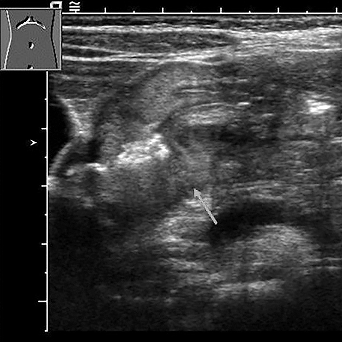
Fig. 2 Transverse gray scale sonogram through the duodenal bulb demonstrating hyperechoic center surrounded by a hypoechoic halo with various thicknesses (arrow).
Reference
-
1. Sung JJ, Kuipers EJ, El-Serag HB. Systematic review: the global incidence and prevalence of peptic ulcer disease. Aliment Pharmacol Ther. 2009; 29:938–946.
Article2. Lanas A, Chan FKL. Peptic ulcer disease. Lancet. 2017; 390:613–624.
Article3. Nord KS. Peptic ulcer disease in the pediatric population. Pediatr Clin North Am. 1988; 35:117–140.
Article4. Blanchard SS, Czinn SJ. Peptic Ulcer Disease in Children. In : Kliegman R, Stanton BMD, St Geme J, Schor NF, editors. Nelson Textbook of Pediatrics. 20th ed. Philadelphia: Elsevier;2016. p. 1816–1819.5. Hayden CK, Swischuk LE, Rytting JE. Gastric ulcer disease in infants: US findings. Radiology. 1987; 164:131–134.
Article6. Herliczek TW, Raghavan D, McCarten K, Wallach M. Sonographic upper gastrointestinal series in the vomiting infant: how we do it. J Clin Imaging Sci. 2011; 1:19.
Article7. Gritzmann N, Hollerweger A, Macheiner P, Rettenbacher T. Transabdominal sonography of the gastrointestinal tract. Eur Radiol. 2002; 12:1748–1761.
Article8. Lim JH, Jeong YM. Sonography of the stomach: an in vitro study to determine the anatomic cause of inner hyperechoic and hypoechoic layers of the gastric wall. AJR Am J Roentgenol. 1994; 162:335–338.
Article9. Lim JH, Lee DH, Ko YT. Sonographic detection of duodenal ulcer. J Ultrasound Med. 1992; 11:91–94.
Article10. Lorentzen T, Nolsøe CP, Khattar SC, Torp-Pedersen ST, Holm HH. Gastric and duodenal wall thickening on abdominal ultrasonography. Positive predictive value. J Ultrasound Med. 1993; 12:633–637.
Article11. Swenson DW, Wallach M. Helicobacter pylori-associated antral gastritis and ulcer disease: imaging by computed tomography and ultrasound. Ultrasound Q. 2012; 28:185–187.12. Horton KM, Fishman EK. Current role of CT in imaging of the stomach. Radiographics. 2003; 23:75–87.
Article13. Sulowski C, Doria AS, Langer JC, Man C, Stephens D, Schuh S. Clinical outcomes in obese and normal-weight children undergoing ultrasound for suspected appendicitis. Acad Emerg Med. 2011; 18:167–173.
Article14. Hörmann M, Scharitzer M, Stadler A, Pokieser P, Puig S, Helbich T. Ultrasound of the appendix in children: is the child too obese? Eur Radiol. 2003; 13:1428–1431.
Article15. Josephson T, Styrud J, Eriksson S. Ultrasonography in acute appendicitis. Body mass index as selection factor for US examination. Acta Radiol. 2000; 41:486–488.
Article16. Yu SH, Kim CB, Park JW, Kim MS, Radosevich DM. Ultrasonography in the diagnosis of appendicitis: evaluation by meta-analysis. Korean J Radiol. 2005; 6:267–277.
Article17. Kotagal M, Richards MK, Flum DR, Acierno SP, Weinsheimer RL, Goldin AB. Use and accuracy of diagnostic imaging in the evaluation of pediatric appendicitis. J Pediatr Surg. 2015; 50:642–646.
Article18. Parente F, Greco S, Molteni M, Cucino C, Maconi G, Sampietro GM, et al. Role of early ultrasound in detecting inflammatory intestinal disorders and identifying their anatomical location within the bowel. Aliment Pharmacol Ther. 2003; 18:1009–1016.
Article19. Hollerbach S, Geissler A, Schiegl H, Kullmann F, Lock G, Schmidt J, et al. The accuracy of abdominal ultrasound in the assessment of bowel disorders. Scand J Gastroenterol. 1998; 33:1201–1208.
Article20. Joharjy IA, Mustafa MA, Zaidi AJ. Fluid-aided sonography of the stomach and duodenum in the diagnosis of peptic ulcer disease in adult patients. J Ultrasound Med. 1990; 9:77–84.
Article21. Liu Z, Guo J, Wang S, Zhao Y, Liu Z, Li J, et al. Evaluation of transabdominal ultrasound with oral cellulose-based contrast agent in the detection and surveillance of gastric ulcer. Ultrasound Med Biol. 2017; 43:1364–1371.
Article
