J Korean Assoc Oral Maxillofac Surg.
2018 Oct;44(5):207-211. 10.5125/jkaoms.2018.44.5.207.
Complications after craniofacial reconstruction with calcium phosphate cements: a case report and review of the literature
- Affiliations
-
- 1Department of Oral and Maxillofacial Surgery, Taleghani Hospital, School of Dentistry, Shahid Beheshti University of Medical Sciences, Tehran, Iran. dr.f.latifi@gmail.com
- 2Department of Plastic and Reconstructive Surgery, Hazrate Fatemeh Hospital, School of Medicine, Iran University of Medical Sciences, Tehran, Iran.
- KMID: 2430580
- DOI: http://doi.org/10.5125/jkaoms.2018.44.5.207
Abstract
- Among different graft materials for craniofacial reconstruction, calcium phosphate cements have the advantages of alloplastic grafts and wide use. The authors report a case of foreign body reaction following frontal reconstruction with JectOS (an injectable calcium orthophosphate cement; Kasios) and reviewed the literature on complications of this material after craniofacial reconstruction from 2002 to 2017. Complications were categorized into two groups: immunologic reactions (consisting of seroma collection, chronic sinus mucosa swelling, and foreign body reaction) and non-immune events (infection, fragmentation, and ejection). It is wise to use calcium phosphate-based material only in selected cases with small defects, and long-term follow-up is needed to observe their consequences.
MeSH Terms
Figure
-
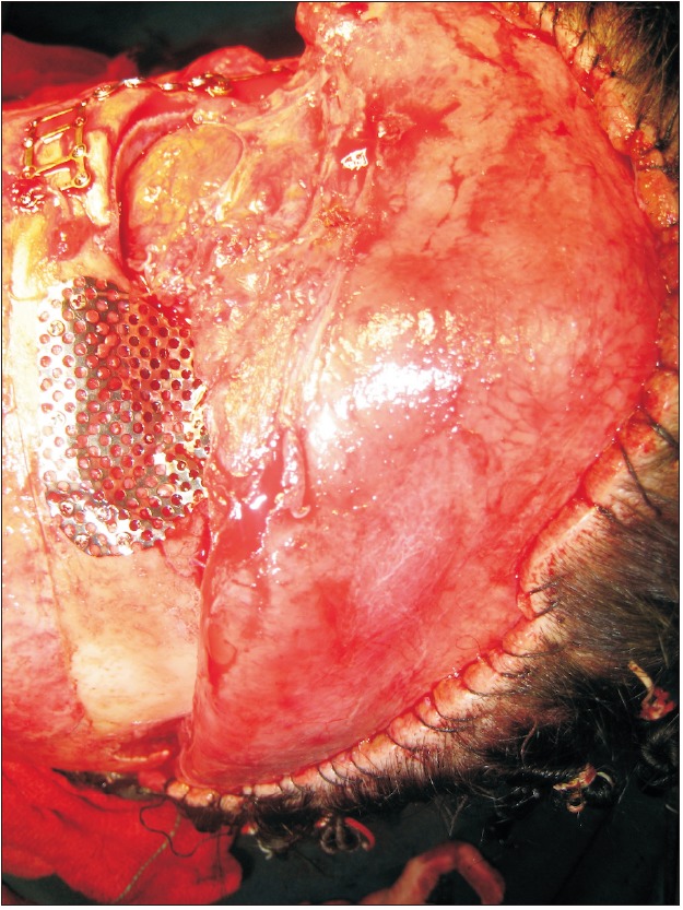
Fig. 1 Reconstruction of the frontal defect with titanium mesh.
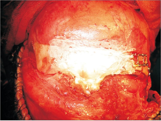
Fig. 2 Coverage of the frontal defect and titanium mesh with JectOS (Kasios).
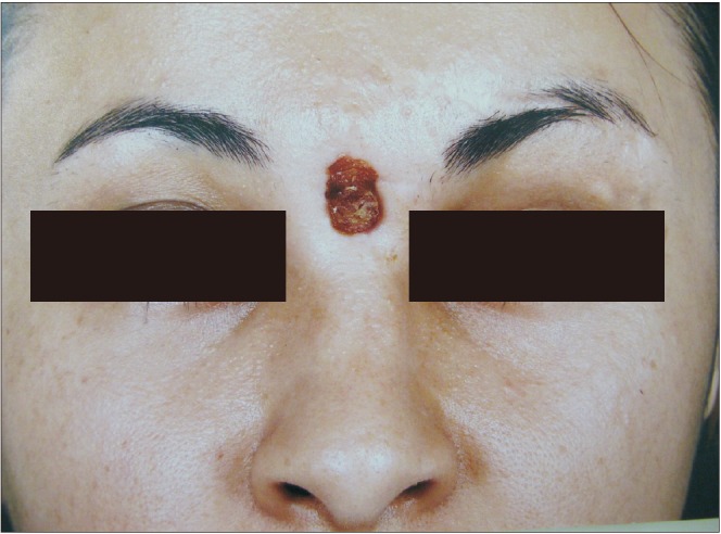
Fig. 3 Midfrontal fistula.
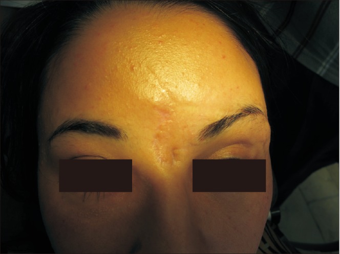
Fig. 4 Postoperation course after debridement and removal of the reconstructive titanium mesh and JectOS (Kasios).
Reference
-
1. Neumann A, Kevenhoerster K. Biomaterials for craniofacial reconstruction. GMS Curr Top Otorhinolaryngol Head Neck Surg. 2009; 8:Doc08. PMID: 22073101.2. Rodriguez ED, Stanwix MG, Nam AJ, St Hilaire H, Simmons OP, Christy MR, et al. Twenty-six-year experience treating frontal sinus fractures: a novel algorithm based on anatomical fracture pattern and failure of conventional techniques. Plast Reconstr Surg. 2008; 122:1850–1866. PMID: 19050539.
Article3. Giannoudis PV, Dinopoulos H, Tsiridis E. Bone substitutes: an update. Injury. 2005; 36(Suppl 3):S20–S27. PMID: 16188545.
Article4. Dorozhkin SV. Self-setting calcium orthophosphate formulations: cements, concretes, pastes and putties. Int J Mat Chem. 2011; 1:1–48.
Article5. Neamat A, Gawish A, Gamal-Eldeen AM. Beta-tricalcium phosphate promotes cell proliferation, osteogenesis and bone regeneration in intrabony defects in dogs. Arch Oral Biol. 2009; 54:1083–1090. PMID: 19828137.6. Ooms EM, Wolke JG, van de Heuvel MT, Jeschke B, Jansen JA. Histological evaluation of the bone response to calcium phosphate cement implanted in cortical bone. Biomaterials. 2003; 24:989–1000. PMID: 12504521.
Article7. Maier W. Biomaterials in skull base surgery. GMS Curr Top Otorhinolaryngol Head Neck Surg. 2009; 8:Doc07. PMID: 22073100.8. Obert L, Lepage D, Gasse N, Rochet S, Garbuio P. Extra-articular distal radius malunion: the phosphate cement alternative. Orthop Traumatol Surg Res. 2010; 96:574–578. PMID: 20634164.
Article9. Nilsson M, Wang JS, Wielanek L, Tanner KE, Lidgren L. Biodegradation and biocompatability of a calcium sulphate-hydroxyapatite bone substitute. J Bone Joint Surg Br. 2004; 86:120–125. PMID: 14765879.
Article10. Chun BD, Kim SW, Lee ST, Kim TH, Lee JH, Kim GC, et al. Interaction between odontoblast and bio-calcium phosphate cement reinforced with chitosan. J Korean Assoc Oral Maxillofac Surg. 2011; 37:415–420.
Article11. Hönig JF, Merten HA, Nitsch A, Verheggen R. Contouring of cranial vault irregularities with hydroxyapatite cement: a clinical and experimental investigation. J Craniofac Surg. 2005; 16:457–460. PMID: 15915115.
Article12. Matic D, Phillips JH. A contraindication for the use of hydroxyapatite cement in the pediatric population. Plast Reconstr Surg. 2002; 110:1–5. PMID: 12087221.
Article13. Magee WP Jr, Ajkay N, Freda N, Rosenblum RS. Use of fast-setting hydroxyapatite cement for secondary craniofacial contouring. Plast Reconstr Surg. 2004; 114:289–297. PMID: 15277791.
Article14. Verret DJ, Ducic Y, Oxford L, Smith J. Hydroxyapatite cement in craniofacial reconstruction. Otolaryngol Head Neck Surg. 2005; 133:897–899. PMID: 16360510.
Article15. Mathur KK, Tatum SA, Kellman RM. Carbonated apatite and hydroxyapatite in craniofacial reconstruction. Arch Facial Plast Surg. 2003; 5:379–383. PMID: 12975134.
Article16. Eppley BL, Hollier L, Stal S. Hydroxyapatite cranioplasty: 2. Clinical experience with a new quick-setting material. J Craniofac Surg. 2003; 14:209–214. PMID: 12621292.
Article17. Durham SR, McComb JG, Levy ML. Correction of large (>25 cm(2)) cranial defects with “reinforced” hydroxyapatite cement: technique and complications. Neurosurgery. 2003; 52:842–845. discussion 845. PMID: 12657179.
Article18. Kerr RG, Hearst MJ, Samy RN, van Loveren HR, Tew JM Jr, Pensak ML, et al. Delayed extrusion of hydroxyapatite cement after transpetrosal reconstruction. Neurosurgery. 2009; 64:527–531. discussion 531-2. PMID: 19240615.
Article19. David L, Argenta L, Fisher D. Hydroxyapatite cement in pediatric craniofacial reconstruction. J Craniofac Surg. 2005; 16:129–133. PMID: 15699660.
Article20. Gómez E, Martín M, Arias J, Carceller F. Clinical applications of Norian SRS (calcium phosphate cement) in craniofacial reconstruction in children: our experience at Hospital La Paz since 2001. J Oral Maxillofac Surg. 2005; 63:8–14. PMID: 15635550.
Article21. Gosain AK, Chim H, Arneja JS. Application-specific selection of biomaterials for pediatric craniofacial reconstruction: developing a rational approach to guide clinical use. Plast Reconstr Surg. 2009; 123:319–330. PMID: 19116568.
Article22. Greenberg BM, Schneider SJ. Alloplastic reconstruction of large cranio-orbital defects: a comparative evaluation. Ann Plast Surg. 2005; 55:43–51. discussion 51. PMID: 15985790.23. Gilardino MS, Cabiling DS, Bartlett SP. Long-term follow-up experience with carbonated calcium phosphate cement (Norian) for cranioplasty in children and adults. Plast Reconstr Surg. 2009; 123:983–994. PMID: 19319064.
Article24. Singh KA, Burstein FD, Williams JK. Use of hydroxyapatite cement in pediatric craniofacial reconstructive surgery: strategies for avoiding complications. J Craniofac Surg. 2010; 21:1130–1135. PMID: 20613591.25. Zins JE, Moreira-Gonzalez A, Papay FA. Use of calcium-based bone cements in the repair of large, full-thickness cranial defects: a caution. Plast Reconstr Surg. 2007; 120:1332–1342. PMID: 17898609.
Article26. Baker SB, Weinzweig J, Kirschner RE, Bartlett SP. Applications of a new carbonated calcium phosphate bone cement: early experience in pediatric and adult craniofacial reconstruction. Plast Reconstr Surg. 2002; 109:1789–1796. PMID: 11994575.
Article27. Anderson JM, Rodriguez A, Chang DT. Foreign body reaction to biomaterials. Semin Immunol. 2008; 20:86–100. PMID: 18162407.
Article28. Zins JE, Langevin CJ, Nasir S. Controversies in skull reconstruction. J Craniofac Surg. 2010; 21:1755–1760. PMID: 21119415.
Article
- Full Text Links
-
- Actions
-
Cited
- CITED
-
- Close
- Share
-
- Similar articles
-
- Clinical Application of the Calcium Phosphate Cement (PolyBone(R)) for the Skull Bone Defects after Microvascular Decompression
- A Case of Acute Phosphate Nephropathy after Sodium Phosphate Preparation
- Lytic Complications after Skull Reconstruction Using GeneX(R)
- The experimental study of the effect of zinc phosphate cement on the solubility of enamel
- Comparative studies on the retentive values of various dental cements used to retain orthodontic bands
