Clin Endosc.
2018 Nov;51(6):596-599. 10.5946/ce.2018.057.
Treatment Using the SpyGlass Digital System in a Patient with Hepatolithiasis after a Whipple Procedure
- Affiliations
-
- 1Department of Gastroenterology, Yamaguchi University Graduate School of Medicine, Ube, Yamaguchi, Japan. harima@yamaguchi-u.ac.jp
- 2Department of Cancer Screening, Ube Industries Central Hospital, Ube, Yamaguchi, Yamaguchi University Graduate School of Medicine, Ube, Yamaguchi, Japan.
- 3Department of Gastroenterology and Hepatology, Yamaguchi University Graduate School of Medicine, Ube, Yamaguchi, Japan.
- KMID: 2430405
- DOI: http://doi.org/10.5946/ce.2018.057
Abstract
- An 89-year-old man was referred to our hospital for treatment of hepatolithiasis causing recurrent cholangitis. He had undergone a prior Whipple procedure. Computed tomography demonstrated left-sided hepatolithiasis. First, we conducted peroral direct cholangioscopy (PDCS) using an ultraslim endoscope. Although PDCS was successfully conducted, it was unsuccessful in removing all the stones. The stones located in the B2 segment were difficult to remove because the endoscope could not be inserted deeply into this segment due to the small size of the intrahepatic bile duct. Next, we substituted the endoscope with an upper gastrointestinal endoscope. After positioning the endoscope, the SpyGlass digital system (SPY-DS) was successfully inserted deep into the B2 segment. Upon visualizing the residual stones, we conducted SPY-DS-guided electrohydraulic lithotripsy. The stones were disintegrated and completely removed. In cases of PDCS failure, a treatment strategy using the SPY-DS can be considered for patients with hepatolithiasis after a Whipple procedure.
MeSH Terms
Figure
-
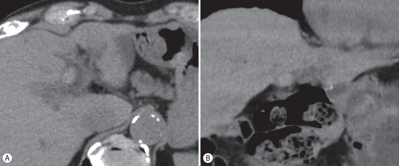
Fig. 1. Computed tomography results. (A) Axial view. (B) Coronal view. Computed tomography images showing a calcified stone impacted within the intrahepatic bile duct of the left lobe.
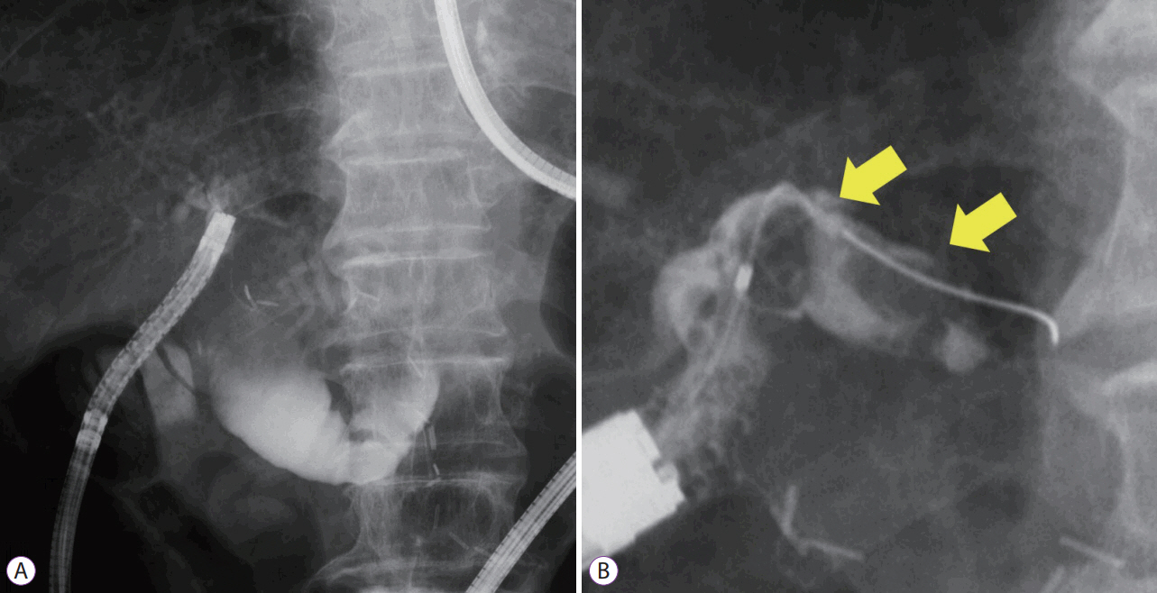
Fig. 2. Cholangiography results. (A) An ultraslim endoscope was inserted directly into the left hepatic duct. (B) Cholangiography image showing two large stones in the common trunk of the intrahepatic bile duct of segment 2 and segment 3 (arrows).
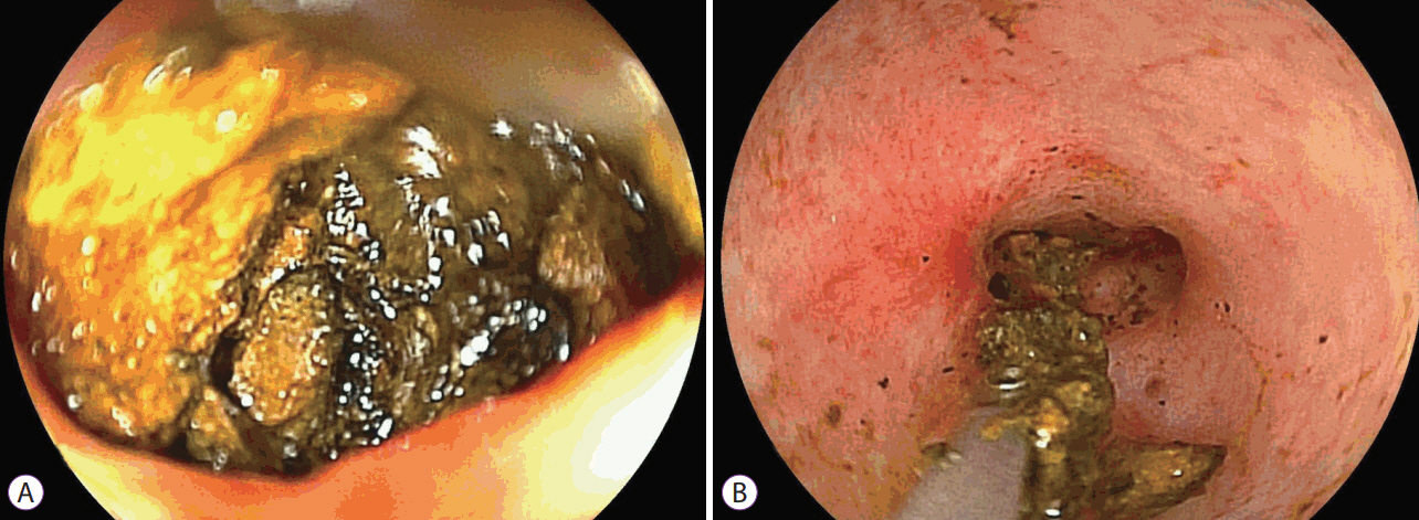
Fig. 3. Peroral direct cholangioscopy (PDCS) results. (A) PDCS image showing a large stone located in the common trunk of the intrahepatic bile duct of segment 2 and segment 3. (B) The volume of the stone was reduced using biopsy forceps during PDCS.
Reference
-
1. Cheon YK, Cho YD, Moon JH, Lee JS, Shim CS. Evaluation of long-term results and recurrent factors after operative and nonoperative treatment for hepatolithiasis. Surgery. 2009; 146:843–853.
Article2. Okugawa T, Tsuyuguchi T, K C S, et al. Peroral cholangioscopic treatment of hepatolithiasis: long-term results. Gastrointest Endosc. 2002; 56:366–371.
Article3. Itoi T, Sofuni A, Itokawa F, et al. Diagnostic and therapeutic peroral direct cholangioscopy in patients with altered GI anatomy (with videos). Gastrointest Endosc. 2012; 75:441–449.
Article4. Matsumoto K, Tsutsumi K, Kato H, et al. Effectiveness of peroral direct cholangioscopy using an ultraslim endoscope for the treatment of hepatolithiasis in patients with hepaticojejunostomy (with video). Surg Endosc. 2016; 30:1249–1254.
Article5. Ghersi S, Fuccio L, Bassi M, Fabbri C, Cennamo V. Current status of peroral cholangioscopy in biliary tract diseases. World J Gastrointest Endosc. 2015; 7:510–517.
Article6. Xu MM, Kahaleh M. Recent developments in choledochoscopy: technical and clinical advances. Clin Exp Gastroenterol. 2016; 9:119–124.7. Parsi MA. Peroral cholangioscopy in the new millennium. World J Gastroenterol. 2011; 17:1–6.
Article8. Chen YK, Pleskow DK. SpyGlass single-operator peroral cholangiopancreatoscopy system for the diagnosis and therapy of bile-duct disorders: a clinical feasibility study (with video). Gastrointest Endosc. 2007; 65:832–841.
Article9. Franzini TA, Moura RN, de Moura EG. Advances in therapeutic cholangioscopy. Gastroenterol Res Pract. 2016; 2016:5249152.
Article10. Ogura T, Imanishi M, Kurisu Y, et al. Prospective evaluation of digital single-operator cholangioscope for diagnostic and therapeutic procedures (with videos). Dig Endosc. 2017; 29:782–789.
Article11. Shimatani M, Matsushita M, Takaoka M, et al. Effective “short” double-balloon enteroscope for diagnostic and therapeutic ERCP in patients with altered gastrointestinal anatomy: a large case series. Endoscopy. 2009; 41:849–854.
Article12. Iwai T, Kida M, Yamauchi H, Imaizumi H, Koizumi W. Short-type and conventional single-balloon enteroscopes for endoscopic retrograde cholangiopancreatography in patients with surgically altered anatomy: single-center experience. Dig Endosc. 2014; 26 Suppl 2:156–163.
Article13. Trikudanathan G, Navaneethan U, Parsi MA. Endoscopic management of difficult common bile duct stones. World J Gastroenterol. 2013; 19:165–173.
Article14. Kamiyama R, Ogura T, Okuda A, et al. Electrohydraulic lithotripsy for difficult bile duct stones under endoscopic retrograde cholangiopancreatography and peroral transluminal cholangioscopy guidance. Gut Liver. 2018; Feb. 8. [Epub]. https://doi.org/10.5009/gnl17352.
Article15. Iwashita T, Doi S, Yasuda I. Endoscopic ultrasound-guided biliary drainage: a review. Clin J Gastroenterol. 2014; 7:94–102.
Article
