A Rare Duodenal Subepithelial Tumor: Duodenal Schwannoma
- Affiliations
-
- 1Department of Internal Medicine, Pusan National University School of Medicine and Biomedical Research Institute, Pusan National University Hospital, Busan, Korea. doc0224@pusan.ac.kr
- 2Department of Pathology, Pusan National University School of Medicine and Biomedical Research Institute, Pusan National University Hospital, Busan, Korea.
- KMID: 2430403
- DOI: http://doi.org/10.5946/ce.2018.050
Abstract
- Schwannomas are uncommon neoplasms that arise from Schwann cells of the neural sheath. Gastrointestinal schwannomas are rare among mesenchymal tumors of the gastrointestinal tract, and only a few cases have been reported to date. Duodenal schwannomas are usually discovered incidentally and achieving a preoperative diagnosis is difficult. Schwannomas can be distinguished from other subepithelial tumors on endoscopic ultrasonography; however, any typical endosonographic features of duodenal schwannomas have not been reported due to the rarity of these tumors. Immunohistochemistry is essential to distinguish schwannomas from gastrointestinal stromal tumors and leiomyomas. We report a case of duodenal schwannoma found incidentally during a health check-up endoscopy. On endoscopic ultrasonography, this tumor was suspected as a gastrointestinal stromal tumor; therefore, the patient underwent laparoscopic wedge resection of the tumor. Histopathology and immunohistochemistry confirmed that the duodenal lesion was a benign schwannoma.
Keyword
MeSH Terms
Figure
-
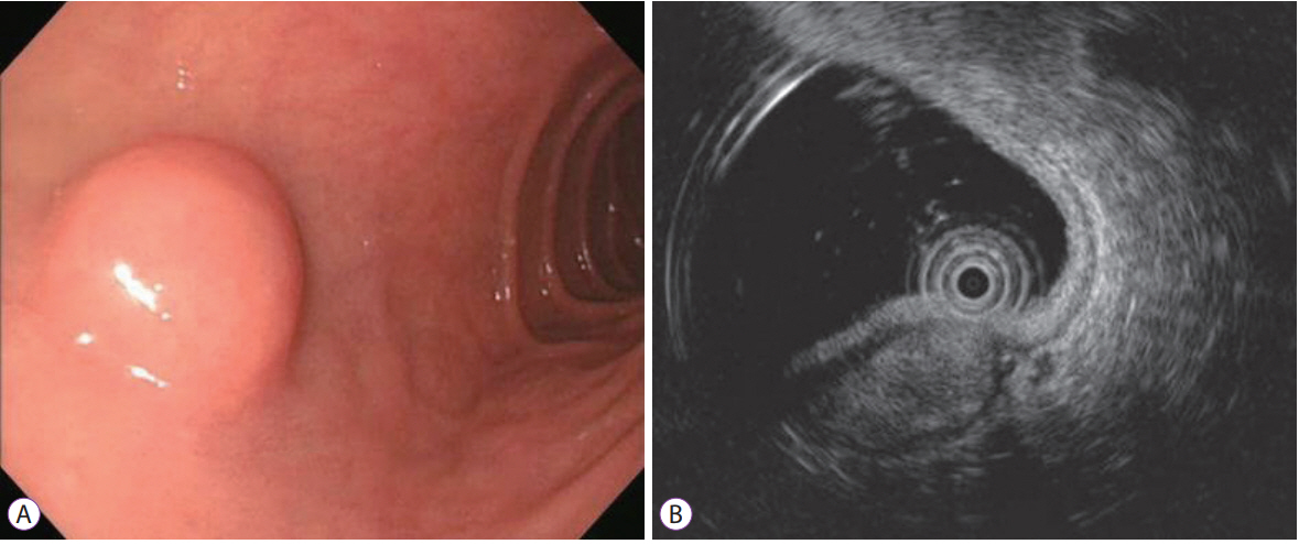
Fig. 1. (A) Upper endoscopy reveals a subepithelial mass covered by the normal mucosa on the duodenal bulb. (B) On endoscopic ultrasonography, the mass is a 1.5×0.7 cm sized heterogeneously hypoechoic mass with marginal haloes originating from the muscularis propria layer.
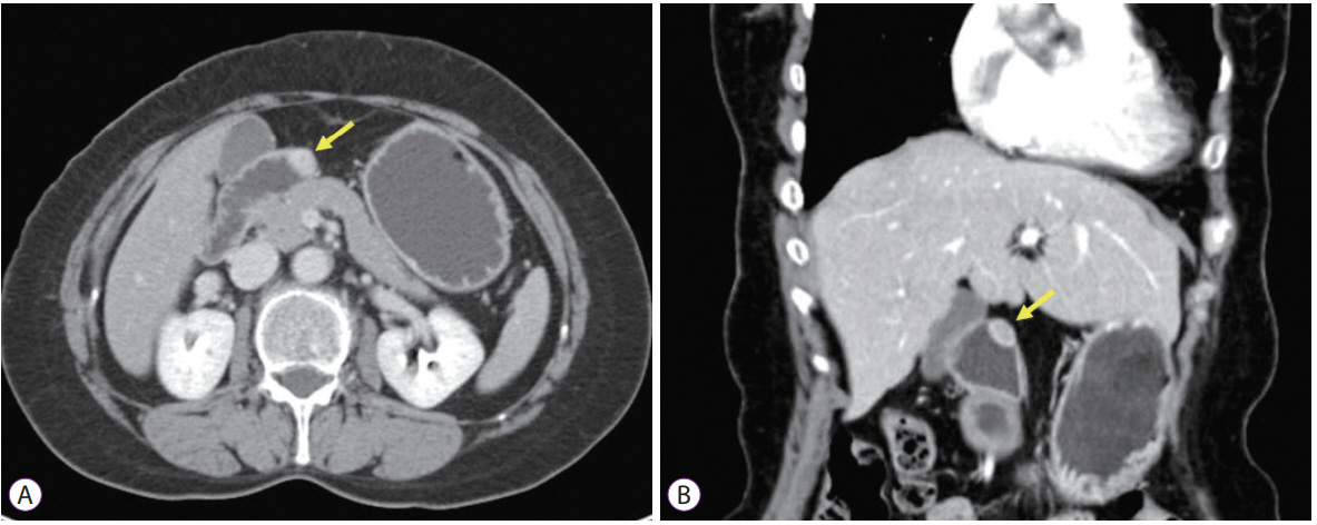
Fig. 2. Abdominopelvic computed tomography scans show a 1.5×1.0 cm sized homogenously-enhancing mass in the anterior wall of the duodenal bulb (arrow). (A) Axial section view. (B) Coronal section view.

Fig. 3. (A) The resected tumor is an intramural, solid, yellowish-to-white mass on cross section. (B) Microscopically, the mass is composed of spindle cells with wavy nuclei. Mild nuclear atypia are seen, but the mitosis count is less than 5/50 high-power fields (hematoxylin and eosin stain, ×200). Immunohistochemically, the tumor cells are strongly positive for S-100 protein, ×400 (C), but negative for smooth muscle actin, ×400 (D), CD34, ×400 (E), and c-kit, ×400 (F).
Reference
-
1. Hou YY, Tan YS, Xu JF, et al. Schwannoma of the gastrointestinal tract: a clinicopathological, immunohistochemical and ultrastructural study of 33 cases. Histopathology. 2006; 48:536–545.
Article2. Miettinen M, Shekitka KM, Sobin LH. Schwannomas in the colon and rectum: a clinicopathologic and immunohistochemical study of 20 cases. Am J Surg Pathol. 2001; 25:846–855.3. Daimaru Y, Kido H, Hashimoto H, Enjoji M. Benign schwannoma of the gastrointestinal tract: a clinicopathologic and immunohistochemical study. Hum Pathol. 1988; 19:257–264.
Article4. Seno K, Itoh M, Endoh K, et al. Schwannoma of the duodenum causing melena. Intern Med. 1994; 33:621–623.5. Meynen P, Van Holsbeeck B, Seynaeve P, Mortelmans L. Duodenal schwannoma. Rofo. 1989; 151:621–622.
Article6. Nilsson B, Jonsson I. Malignant neurinoma of the duodenum; report of one case and review of the literature. Acta Chir Scand. 1957; 113:357–363.7. Woodruff JM, Selig AM, Crowley K, Allen PW. Schwannoma (neurilemoma) with malignant transformation. A rare, distinctive peripheral nerve tumor. Am J Surg Pathol. 1994; 18:882–895.
Article8. Kazerooni EA, Quint LE, Francis IR. Duodenal neoplasms: predictive value of CT for determining malignancy and tumor resectability. AJR Am J Roentgenol. 1992; 159:303–309.
Article9. Kim GH, Cho YK, Kim EY, et al. Comparison of 22-gauge aspiration needle with 22-gauge biopsy needle in endoscopic ultrasonography-guided subepithelial tumor sampling. Scand J Gastroenterol. 2014; 49:347–354.
Article10. Yoon JM, Kim GH, Park DY, et al. Endosonographic features of gastric schwannoma: a single center experience. Clin Endosc. 2016; 49:548–554.
Article11. Park HC, Son DJ, Oh HH, et al. Endoscopic ultrasonographic characteristics of gastric schwannoma distinguished from gastrointestinal stromal tumor. Korean J Gastroenterol. 2015; 65:21–26.
Article12. Jung MK, Jeon SW, Cho CM, et al. Gastric schwannomas: endosonographic characteristics. Abdom Imaging. 2008; 33:388–390.
Article13. Levy AD, Quiles AM, Miettinen M, Sobin LH. Gastrointestinal schwannomas: CT features with clinicopathologic correlation. AJR Am J Roentgenol. 2005; 184:797–802.
Article14. Takeda M, Amano Y, Machida T, Kato S, Naito Z, Kumita S. CT, MRI, and PET findings of gastric schwannoma. Jpn J Radiol. 2012; 30:602–605.
Article
- Full Text Links
-
- Actions
-
Cited
- CITED
-
- Close
- Share
-
- Similar articles
-
- Annular Pancreas with a Duodenal Web: a Rare Presentation with Simultaneous Intrinsic and Extrinsic Duodenal Obstruction
- A Case of Resolution of Duodenal Obstruction Caused by Duodenal Ulcer after the Eradication of Helicobacter pylori Infection
- Three Cases of Coexistence of Gastric Cancer and Duodenal Ulcer
- Gastroscopic Observation of Duodenal Tuberculosis
- A Case of Primary Aortoenteric Fistula Mimicking Duodenal Subepithelial Tumor
