Role of Myocardial Extracellular Volume Fraction Measured with Magnetic Resonance Imaging in the Prediction of Left Ventricular Functional Outcome after Revascularization of Chronic Total Occlusion of Coronary Arteries
- Affiliations
-
- 1Department of Radiology, Zhongshan Hospital, Fudan University; Department of Medical Imaging, Shanghai Medical School, Fudan University and Shanghai Institute of Medical Imaging, Shanghai, China. zengmengsu@outlook.com
- 2Department of Nuclear Medicine, The First Affiliated Hospital of Soochow University, Suzhou, China.
- 3Department of Cardiology, Zhongshan Hospital, Fudan University, Shanghai, China.
- 4Siemens Healthcare GmbH, Erlangen, Germany.
- 5Siemens Shenzhen Magnetic Resonance (C.F.), Shenzhen, China.
- 6Department of Clinical Laboratory, Zhongshan Hospital, Fudan University, Shanghai, China.
- KMID: 2429922
- DOI: http://doi.org/10.3348/kjr.2018.0069
Abstract
OBJECTIVE
The purpose of this study was to prospectively investigate the value of the myocardial extracellular volume fraction (ECV) in predicting myocardial functional outcome after revascularization of coronary chronic total occlusion (CTO).
MATERIALS AND METHODS
Thirty patients with CTO underwent cardiovascular magnetic resonance (CMR) before and 6 months after revascularization. Three baseline markers of functional outcome were evaluated in the dysfunctional segments assigned to the CTO vessels: ECV, transmural extent of infarction (TEI), and unenhanced rim thickness (RIM). At the global level, the ECV values of the whole myocardium with and without a hyperenhanced region (global and remote ECV) were respectively measured.
RESULTS
In per-segment analysis, ECV was superior to TEI and RIM in predicting functional recovery (area under receiver operating characteristic curve [AUC]: 0.86 vs. 0.75 and 0.73, all p values < 0.010), and it emerged as the only independent predictor of regional functional outcome (odds ratio [OR] = 0.83, 95% confidence interval [CI]: 0.77-0.89; p < 0.001) independent of collateral circulation. In per-patient analysis, global baseline ECV was indicative of ejection fraction (EF) at the follow-up examination (β = −0.61, p < 0.001) and changes in EF (β = −0.57, p = 0.001) in multivariate regression analysis. A patient with global baseline ECV less than 30.0% (AUC, 0.93; sensitivity 94%, specificity 80%) was more likely to demonstrate significant EF improvement (OR: 0.38; 95% CI: 0.17-0.85; p = 0.019).
CONCLUSION
Extracellular volume fraction obtained by CMR may provide incremental value for the prediction of functional recovery both at the segmental and global levels in CTO patients, and may facilitate the identification of patients who can benefit from revascularization.
Keyword
MeSH Terms
Figure
-
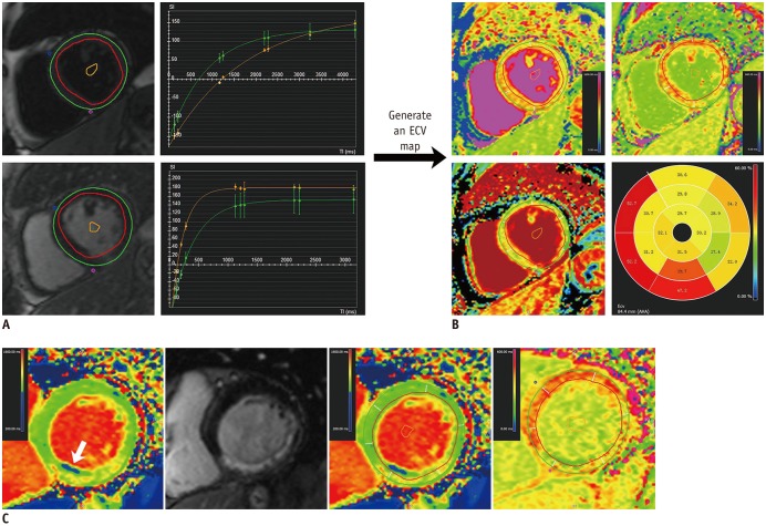
Fig. 1 Measurement of ECV based on AHA 16-segment model in patients with right coronary artery chronic total occlusion.A. “T1 Calculation” module required endocardial and epicardial contours, segmentation points and blood-pool contour in all slices in both T1 maps. B. “Map Analysis” module generated ECV map and polar map when hematocrit was entered and long axis extent was defined. Note that AHA 2 and 3 segments were located at outflow tract. C. In another case with chronic fatty infiltration (arrow), contours were drawn avoiding zone with abnormal low native T1 times, and copied to matched post-contrast T1 map. AHA = American Heart Association, ECV = extracellular volume fraction
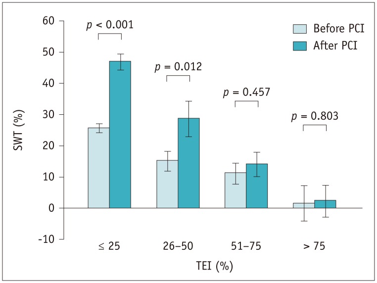
Fig. 2 Bar chart reveals inverse correlation between TEI and improvement in SWT.Bars and error bars represent means and 1 standard error of mean respectively. PCI = percutaneous coronary intervention, SWT = segmental wall thickening, TEI = transmural extent of infarction
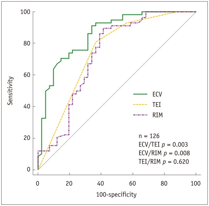
Fig. 3 ROC curves demonstrating diagnostic performance of baseline imaging markers in predicting segmental function recovery.RIM = unenhanced rim thickness, ROC = receive operating characteristic
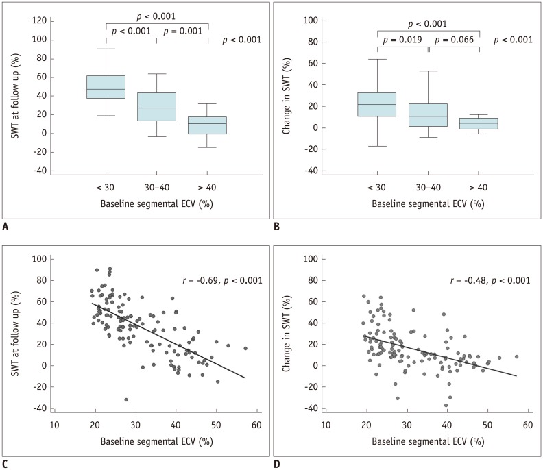
Fig. 4 Correlation between baseline segmental ECV, SWT at follow-up (A, C), and change in SWT from follow-up to baseline (B, D).
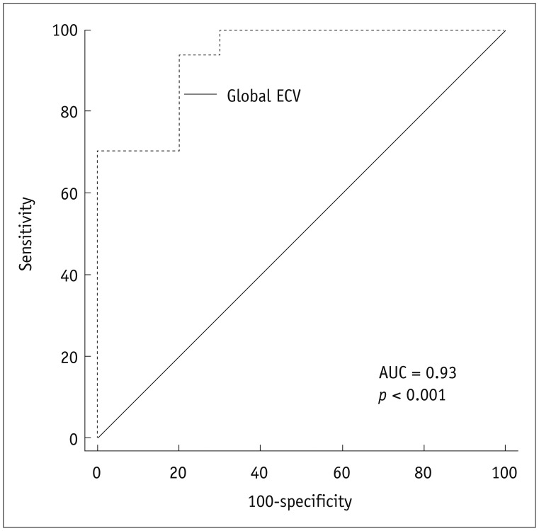
Fig. 5 ROC curves for prediction of significant improvement in ejection fraction based on global ECV at baseline.AUC = area under receiver operating characteristic curve
Cited by 6 articles
-
RE: Prediction of the Left Ventricular Functional Outcome by Myocardial Extracellular Volume Fraction Measured Using Magnetic Resonance Imaging: Methodological Issue
Shiva Karimi, Mojtaba Pourmehdi, Mehdi Naderi
Korean J Radiol. 2019;20(6):1001-1002. doi: 10.3348/kjr.2019.0062.Reply: Prediction of the Left Ventricular Functional Outcome by Myocardial Extracellular Volume Fraction Measured Using Magnetic Resonance Imaging; Methodological Issue
Yinyin Chen, Mengsu Zeng
Korean J Radiol. 2019;20(8):1311-1312. doi: 10.3348/kjr.2019.0115.Guidelines for Cardiovascular Magnetic Resonance Imaging from the Korean Society of Cardiovascular Imaging—Part 2: Interpretation of Cine, Flow, and Angiography Data
Jae Wook Lee, Jee Hye Hur, Dong Hyun Yang, Bae Young Lee, Dong Jin Im, Su Jin Hong, Eun Young Kim, Eun-Ah Park, Yeseul Jo, JeongJae Kim, Chul Hwan Park, Hwan Seok Yong
Korean J Radiol. 2019;20(11):1477-1490. doi: 10.3348/kjr.2019.0407.Guideline for Cardiovascular Magnetic Resonance Imaging from the Korean Society of Cardiovascular Imaging—Part 1: Standardized Protocol
Yeseul Jo, JeongJae Kim, Chul Hwan Park, Jae Wook Lee, Jee Hye Hur, Dong Hyun Yang, Bae Young Lee, Dong Jin Im, Su Jin Hong, Eun Young Kim, Eun-Ah Park, Pan Ki Kim, Hwan Seok Yong
Korean J Radiol. 2019;20(9):1313-1333. doi: 10.3348/kjr.2019.0398.Guidelines for Cardiovascular Magnetic Resonance Imaging from the Korean Society of Cardiovascular Imaging—Part 3: Perfusion, Delayed Enhancement, and T1- and T2 Mapping
Dong Jin Im, Su Jin Hong, Eun-Ah Park, Eun Young Kim, Yeseul Jo, JeongJae Kim, Chul Hwan Park, Hwan Seok Yong, Jae Wook Lee, Jee Hye Hur, Dong Hyun Yang, Bae Young Lee
Korean J Radiol. 2019;20(12):1562-1582. doi: 10.3348/kjr.2019.0411.Guidelines for Cardiovascular Magnetic Resonance Imaging from the Korean Society of Cardiovascular Imaging (KOSCI) - Part 3: Perfusion, Delayed Enhancement, and T1- and T2 Mapping
Dong Jin Im, Su Jin Hong, Eun-Ah Park, Eun Young Kim, Yeseul Jo, Jeong Jae Kim, Chul Hwan Park, Hwan Seok Yong, Jae Wook Lee, Jee Hye Hur, Dong Hyun Yang, Bae-Young Lee
Investig Magn Reson Imaging. 2020;24(1):1-20. doi: 10.13104/imri.2020.24.1.1.
Reference
-
1. Brilakis ES, Karmpaliotis D, Vo MN, Garcia S, Michalis L, Alaswad K, et al. Advances in the management of coronary chronic total occlusions. J Cardiovasc Transl Res. 2014; 7:426–436. PMID: 24634196.
Article2. Christofferson RD, Lehmann KG, Martin GV, Every N, Caldwell JH, Kapadia SR. Effect of chronic total coronary occlusion on treatment strategy. Am J Cardiol. 2005; 95:1088–1091. PMID: 15842978.
Article3. Christakopoulos GE, Christopoulos G, Carlino M, Jeroudi OM, Roesle M, Rangan BV, et al. Meta-analysis of clinical outcomes of patients who underwent percutaneous coronary interventions for chronic total occlusions. Am J Cardiol. 2015; 115:1367–1375. PMID: 25784515.
Article4. Pujadas S, Martin V, Rosselló X, Carreras F, Barros A, Leta R, et al. Improvement of myocardial function and perfusion after successful percutaneous revascularization in patients with chronic total coronary occlusion. Int J Cardiol. 2013; 169:147–152. PMID: 24120215.
Article5. Chadid P, Markovic S, Bernhardt P, Hombach V, Rottbauer W, Wöhrle J. Improvement of regional and global left ventricular function in magnetic resonance imaging after recanalization of true coronary chronic total occlusions. Cardiovasc Revasc Med. 2015; 16:228–232. PMID: 25892532.6. Chen YY, Zhang WG, Yang S, Yun H, Deng SM, Fu CX, et al. Extracellular volume fraction in coronary chronic total occlusion patients. Int J Cardiovasc Imaging. 2015; 31:1211–1121. PMID: 25985941.
Article7. Choi JH, Chang SA, Choi JO, Song YB, Hahn JY, Choi SH, et al. Frequency of myocardial infarction and its relationship to angiographic collateral flow in territories supplied by chronically occluded coronary arteries. Circulation. 2013; 127:703–709. PMID: 23277308.
Article8. Romero J, Xue X, Gonzalez W, Garcia MJ. CMR imaging assessing viability in patients with chronic ventricular dysfunction due to coronary artery disease: a meta-analysis of prospective trials. JACC Cardiovasc Imaging. 2012; 5:494–508. PMID: 22595157.9. Kirschbaum SW, Rossi A, Boersma E, Springeling T, van de Ent M, Krestin GP, et al. Combining magnetic resonance viability variables better predicts improvement of myocardial function prior to percutaneous coronary intervention. Int J Cardiol. 2012; 159:192–197. PMID: 21414675.
Article10. Knuesel PR, Nanz D, Wyss C, Buechi M, Kaufmann PA, von Schulthess GK, et al. Characterization of dysfunctional myocardium by positron emission tomography and magnetic resonance: relation to functional outcome after revascularization. Circulation. 2003; 108:1095–1100. PMID: 12939229.
Article11. Arheden H, Saeed M, Higgins CB, Gao DW, Bremerich J, Wyttenbach R, et al. Measurement of the distribution volume of gadopentetate dimeglumine at echo-planar MR imaging to quantify myocardial infarction: comparison with 99mTc-DTPA autoradiography in rats. Radiology. 1999; 211:698–708. PMID: 10352594.12. Ugander M, Oki AJ, Hsu LY, Kellman P, Greiser A, Aletras AH, et al. Extracellular volume imaging by magnetic resonance imaging provides insights into overt and sub-clinical myocardial pathology. Eur Heart J. 2012; 33:1268–1278. PMID: 22279111.
Article13. Bauner KU, Biffar A, Theisen D, Greiser A, Zech CJ, Nguyen ET, et al. Extracellular volume fractions in chronic myocardial infarction. Invest Radiol. 2012; 47:538–545. PMID: 22836311.
Article14. Kirschbaum SW, Rossi A, van Domburg RT, Gruszczynska K, Krestin GP, Serruys PW, et al. Contractile reserve in segments with nontransmural infarction in chronic dysfunctional myocardium using low-dose dobutamine CMR. JACC Cardiovasc Imaging. 2010; 3:614–622. PMID: 20541717.
Article15. Chen YY, Ren DY, Zeng MS, Yang S, Yun H, Fu CX, et al. Myocardial extracellular volume fraction measurement in chronic total coronary occlusion: association with myocardial injury, angiographic collateral flow, and functional recovery. J Magn Reson Imaging. 2016; 44:972–982. PMID: 27008315.
Article16. Cerqueira MD, Weissman NJ, Dilsizian V, Jacobs AK, Kaul S, Laskey WK, et al. American Heart Association Writing Group on Myocardial Segmentation and Registration for Cardiac Imaging. Standardized myocardial segmentation and nomenclature for tomographic imaging of the heart. A statement for healthcare professionals from the Cardiac Imaging Committee of the Council on Clinical Cardiology of the American Heart Association. Int J Cardiovasc Imaging. 2002; 18:539–542. PMID: 12135124.
Article17. Kim RJ, Wu E, Rafael A, Chen EL, Parker MA, Simonetti O, et al. The use of contrast-enhanced magnetic resonance imaging to identify reversible myocardial dysfunction. N Engl J Med. 2000; 343:1445–1453. PMID: 11078769.
Article18. Rassaf T, Nolte J, Heussen N, Krombach GA, Günther RW, Kelm M, et al. Quantitation of the thickness of the non-enhanced myocardial rim predicts recovery of territorial myocardial function in chronic ischemic heart disease: a cardiac magnetic resonance imaging study. Clin Res Cardiol. 2010; 99:293–300. PMID: 20151141.
Article19. Glaveckaite S, Valeviciene N, Palionis D, Puronaite R, Serpytis P, Laucevicius A. Prediction of long-term segmental and global functional recovery of hibernating myocardium after revascularisation based on low dose dobutamine and late gadolinium enhancement cardiovascular magnetic resonance. J Cardiovasc Magn Reson. 2014; 16:83. PMID: 25279683.
Article20. Glaveckaite S, Valeviciene N, Palionis D, Skorniakov V, Celutkiene J, Tamosiunas A, et al. Value of scar imaging and inotropic reserve combination for the prediction of segmental and global left ventricular functional recovery after revascularisation. J Cardiovasc Magn Reson. 2011; 13:35. PMID: 21787383.
Article21. Krittayaphong R, Laksanabunsong P, Maneesai A, Saiviroonporn P, Udompunturak S, Chaithiraphan V. Comparison of cardiovascular magnetic resonance of late gadolinium enhancement and diastolic wall thickness to predict recovery of left ventricular function after coronary artery bypass surgery. J Cardiovasc Magn Reson. 2008; 10:41. PMID: 18808697.
Article22. Kirschbaum SW, Baks T, van den Ent M, Sianos G, Krestin GP, Serruys PW, et al. Evaluation of left ventricular function three years after percutaneous recanalization of chronic total coronary occlusions. Am J Cardiol. 2008; 101:179–185. PMID: 18178403.
Article23. Bondarenko O, Beek AM, Twisk JW, Visser CA, van Rossum AC. Time course of functional recovery after revascularization of hibernating myocardium: a contrast-enhanced cardiovascular magnetic resonance study. Eur Heart J. 2008; 29:2000–2005. PMID: 18556713.
Article24. Pegg TJ, Selvanayagam JB, Jennifer J, Francis JM, Karamitsos TD, Dall'Armellina E, et al. Prediction of global left ventricular functional recovery in patients with heart failure undergoing surgical revascularisation, based on late gadolinium enhancement cardiovascular magnetic resonance. J Cardiovasc Magn Reson. 2010; 12:56. PMID: 20929540.
Article25. Baks T, van Geuns RJ, Duncker DJ, Cademartiri F, Mollet NR, Krestin GP, et al. Prediction of left ventricular function after drug-eluting stent implantation for chronic total coronary occlusions. J Am Coll Cardiol. 2006; 47:721–725. PMID: 16487835.
Article26. Fishbein MC, Maclean D, Maroko PR. The histopathologic evolution of myocardial infarction. Chest. 1978; 73:843–849. PMID: 657859.
Article27. Kidambi A, Motwani M, Uddin A, Ripley DP, McDiarmid AK, Swoboda PP, et al. Myocardial extracellular volume estimation by CMR predicts functional recovery following acute MI. JACC Cardiovasc Imaging. 2017; 10:989–999. PMID: 27771398.
Article28. Zhang J, Li Y, Li M, Pan J, Lu Z. Collateral vessel opacification with CT in patients with coronary total occlusion and its relationship with downstream myocardial infarction. Radiology. 2014; 271:703–710. PMID: 24555634.
Article29. Liu D, Borlotti A, Viliani D, Jerosch-Herold M, Alkhalil M, De Maria GL, et al. CMR native T1 mapping allows differentiation of reversible versus irreversible myocardial damage in ST-segment-elevation myocardial infarction: an OxAMI Study (Oxford acute myocardial infarction). Circ Cardiovasc Imaging. 2017; 10:pii: e005986.
Article30. Bucciarelli-Ducci C, Auger D, Di Mario C, Locca D, Petryka J, O'Hanlon R, et al. CMR guidance for recanalization of coronary chronic total occlusion. JACC Cardiovasc Imaging. 2016; 9:547–556. PMID: 27085432.31. Nakachi T, Kato S, Kirigaya H, Iinuma N, Fukui K, Saito N, et al. Prediction of functional recovery after percutaneous coronary revascularization for chronic total occlusion using late gadolinium enhanced magnetic resonance imaging. J Cardiol. 2017; 69:836–842. PMID: 28256296.
Article32. Altiok E, Tiemann S, Becker M, Koos R, Zwicker C, Schroeder J, et al. Myocardial deformation imaging by two-dimensional speckle-tracking echocardiography for prediction of global and segmental functional changes after acute myocardial infarction: a comparison with late gadolinium enhancement cardiac magnetic resonance. J Am Soc Echocardiogr. 2014; 27:249–257. PMID: 24368027.
Article33. Carrick D, Haig C, Rauhalammi S, Ahmed N, Mordi I, McEntegart M, et al. Prognostic significance of infarct core pathology revealed by quantitative non-contrast in comparison with contrast cardiac magnetic resonance imaging in reperfused ST-elevation myocardial infarction survivors. Eur Heart J. 2016; 37:1044–1059. PMID: 26261290.
Article34. Mollema SA, Delgado V, Bertini M, Antoni ML, Boersma E, Holman ER, et al. Viability assessment with global left ventricular longitudinal strain predicts recovery of left ventricular function after acute myocardial infarction. Circ Cardiovasc Imaging. 2010; 3:15–23. PMID: 19820202.
Article35. Wong TC, Piehler K, Meier CG, Testa SM, Klock AM, Aneizi AA, et al. Association between extracellular matrix expansion quantified by cardiovascular magnetic resonance and short-term mortality. Circulation. 2012; 126:1206–1216. PMID: 22851543.
Article36. Bulluck H, Rosmini S, Abdel-Gadir A, White SK, Bhuva AN, Treibel TA, et al. Automated extracellular volume fraction mapping provides insights into the pathophysiology of left ventricular remodeling post-reperfused ST-elevation myocardial infarction. J Am Heart Assoc. 2016; 5:pii: e003555.
Article37. Fefer P, Knudtson ML, Cheema AN, Galbraith PD, Osherov AB, Yalonetsky S, et al. Current perspectives on coronary chronic total occlusions: the Canadian multicenter chronic total occlusions registry. J Am Coll Cardiol. 2012; 59:991–997. PMID: 22402070.
- Full Text Links
-
- Actions
-
Cited
- CITED
-
- Close
- Share
-
- Similar articles
-
- RE: Prediction of the Left Ventricular Functional Outcome by Myocardial Extracellular Volume Fraction Measured Using Magnetic Resonance Imaging: Methodological Issue
- Reply: Prediction of the Left Ventricular Functional Outcome by Myocardial Extracellular Volume Fraction Measured Using Magnetic Resonance Imaging; Methodological Issue
- Dobutamine Echocardiography in Chronic Coronary Artery Disease with Left Ventricular Dysfunction
- Role of Cardiac Computed Tomography in the Diagnosis of Left Ventricular Myocardial Diseases
- Assessment of Myocardial Viability Using PET
