Lobular Breast Carcinoma Metastasis to the Thyroid Gland: Case Report and Literature Review
- Affiliations
-
- 1Department of Medical Oncology, Limoges University Hospital, Limoges, France. elise.deluche@chu-limoges.fr
- 2Department of Pathology, Limoges University Hospital, Limoges, France.
- KMID: 2429826
- DOI: http://doi.org/10.4048/jbc.2018.21.e55
Abstract
- Metastasis from primary cancer to the thyroid is uncommon in breast cancer. Here we present a case of lobular breast carcinoma that metastasized to the thyroid. A 54-year-old woman without symptoms was admitted to our institution for staging of the lymph node above the left clavicle. An ¹â¸F-fluoro-deoxy-D-glucose positron emission tomography scan was performed for staging, and low uptakes were observed in the left supraclavicular and cervical lymph nodes. High uptake was seen in the posterior and lower left lobe of the thyroid. Histologic findings indicated lobular breast carcinoma (positive GATA3, loss of E-cadherin expression) metastatic to the thyroid with a luminal profile. Immunohistochemical analysis was negative for primary thyroid or parathyroid carcinoma. To our knowledge, this is the first report of a patient presenting a metastatic invasive lobular carcinoma in the thyroid and lymph nodes without a prior diagnosis of breast cancer.
Keyword
MeSH Terms
Figure
-
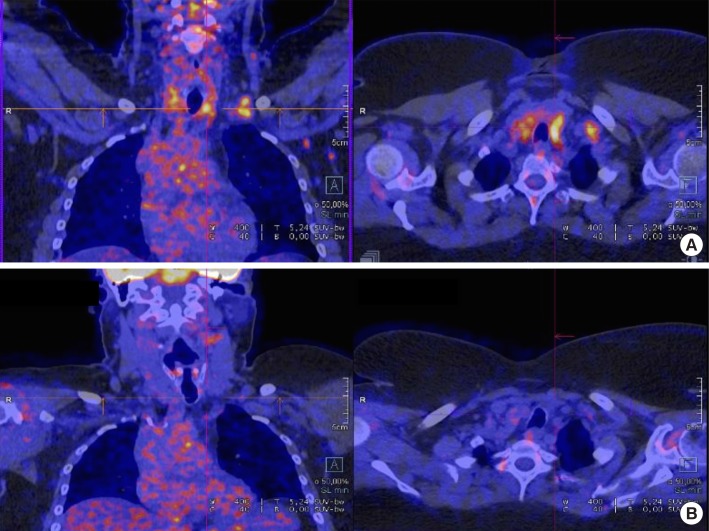
Figure 1 18F-fluoro-deoxy-D-glucose positron emission tomography staging scan for patient with lobular breast carcinoma metastasis to the thyroid gland. (A) April 2016. (B) July 2016.
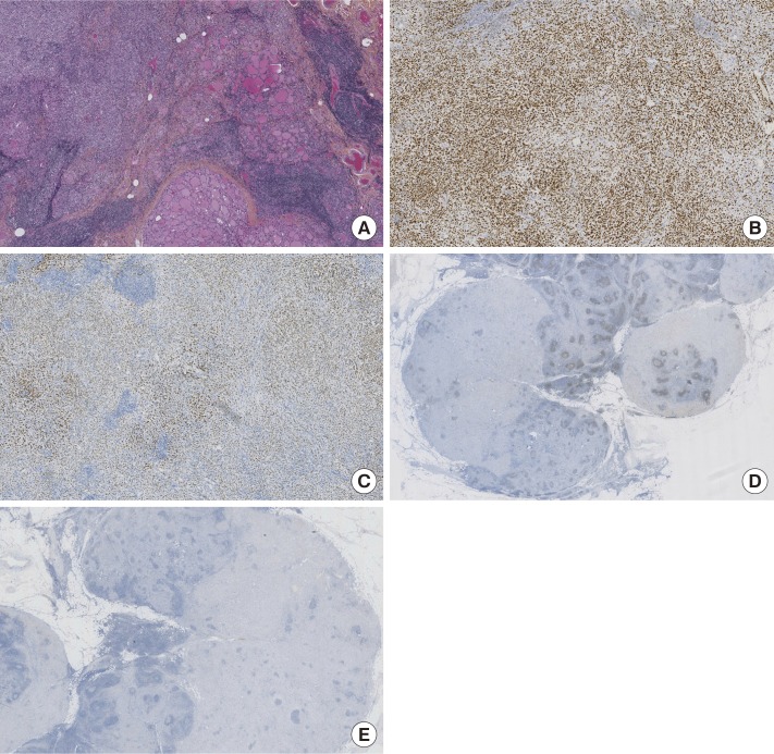
Figure 2 Histologic section from thyroid. Immunohistochemical features were presented. Infiltrating carcinoma which was made up of monomorphic cells without any glandular organization. (A) H&E-stained section of tumor. (B) Immunohistochemical positive staining for GATA3. (C) Immunohistochemical positive staining for estrogen receptor. (D) Immunohistochemical negative staining for PAX8. (E) Immunohistochemical negative staining for thyroid transcription factor 1 (A–E, ×50).
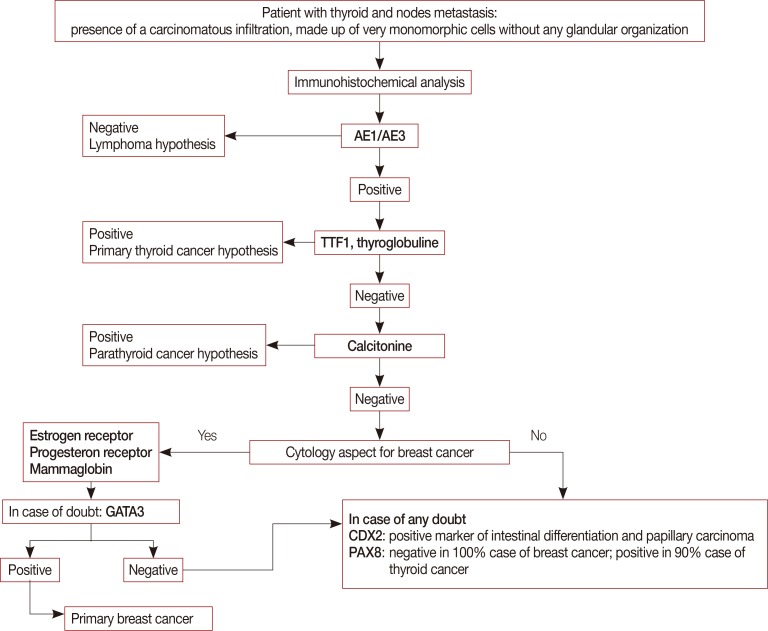
Figure 3 A decision tree to diagnose lobular breast carcinoma metastasis to the thyroid gland.TTF1=thyroid transcription factor 1.
Reference
-
1. Fernández B, Paish EC, Green AR, Lee AH, Macmillan RD, Ellis IO, et al. Lymph-node metastases in invasive lobular carcinoma are different from those in ductal carcinoma of the breast. J Clin Pathol. 2011; 64:995–1000. PMID: 21712309.2. Ferlicot S, Vincent-Salomon A, Médioni J, Genin P, Rosty C, Sigal-Zafrani B, et al. Wide metastatic spreading in infiltrating lobular carcinoma of the breast. Eur J Cancer. 2004; 40:336–341. PMID: 14746850.3. Egaña N, Socias C, Matteucci T, Bilbao I, Alvarez-Coca M. Thyroid metastasis of lobular breast carcinoma. Endocrinol Nutr. 2012; 59:219–220. PMID: 22138624.
Article4. Magers MJ, Dueber JC, Lew M, Pang JC, Davenport RD. Metastatic ductal carcinoma of the breast to the thyroid gland diagnosed with fine needle aspiration: a case report with emphasis on morphologic and immunophenotypic features. Diagn Cytopathol. 2016; 44:530–534. PMID: 26932153.
Article5. Lacka K, Breborowicz D, Uliasz A, Teresiak M. Thyroid metastases from a breast cancer diagnosed by fine-needle aspiration biopsy: case report and overview of the literature. Exp Oncol. 2012; 34:129–133. PMID: 23013767.6. Kim TY, Kim WB, Gong G, Hong SJ, Shong YK. Metastasis to the thyroid diagnosed by fine-needle aspiration biopsy. Clin Endocrinol (Oxf). 2005; 62:236–241. PMID: 15670202.
Article7. Owens CL, Basaria S, Nicol TL. Metastatic breast carcinoma involving the thyroid gland diagnosed by fine-needle aspiration: a case report. Diagn Cytopathol. 2005; 33:110–115. PMID: 16007653.
Article8. Hegerova L, Griebeler ML, Reynolds JP, Henry MR, Gharib H. Metastasis to the thyroid gland: report of a large series from the Mayo Clinic. Am J Clin Oncol. 2015; 38:338–342. PMID: 23799287.9. Cristofanilli M, Gonzalez-Angulo A, Sneige N, Kau SW, Broglio K, Theriault RL, et al. Invasive lobular carcinoma classic type: response to primary chemotherapy and survival outcomes. J Clin Oncol. 2005; 23:41–48. PMID: 15625359.
Article10. Choi YJ, Pinto MM, Hao L, Riba AK. Interobserver variability and aberrant E-cadherin immunostaining of lobular neoplasia and infiltrating lobular carcinoma. Mod Pathol. 2008; 21:1224–1237. PMID: 18587329.
Article11. Cimino-Mathews A, Subhawong AP, Illei PB, Sharma R, Halushka MK, Vang R, et al. GATA3 expression in breast carcinoma: utility in triple-negative, sarcomatoid, and metastatic carcinomas. Hum Pathol. 2013; 44:1341–1349. PMID: 23375642.
Article12. Sujoy V, Pinto A, Nosé V. Columnar cell variant of papillary thyroid carcinoma: a study of 10 cases with emphasis on CDX2 expression. Thyroid. 2013; 23:714–719. PMID: 23488912.
Article13. Laury AR, Perets R, Piao H, Krane JF, Barletta JA, French C, et al. A comprehensive analysis of PAX8 expression in human epithelial tumors. Am J Surg Pathol. 2011; 35:816–826. PMID: 21552115.
Article14. Tacha D, Zhou D, Cheng L. Expression of PAX8 in normal and neoplastic tissues: a comprehensive immunohistochemical study. Appl Immunohistochem Mol Morphol. 2011; 19:293–299. PMID: 21285870.
- Full Text Links
-
- Actions
-
Cited
- CITED
-
- Close
- Share
-
- Similar articles
-
- Nodular Metastatic Carcinoma from Invasive Lobular Breast Cancer
- Metastatic Medullary Carcinoma of Thyroid to Breast; A Case Initially Diagnosed as Primary Invasive Lobular Carcinoma: A Case Report
- Ultrasonographic Features and the Diagnostic Role of Core Needle Biopsy at Metastatic Breast Cancer in the Thyroid gland: A Case Report
- Two Cases of Renal Cell Carcinoma Metastatic to the Thyroid Gland
- A Case of Lymphoepithelioma-Like Carcinoma in the Thyroid Gland
