Prolonged Gallbladder Hydrops in a Kawasaki Disease Patient
- Affiliations
-
- 1Department of Pediatrics, Kyung Hee University Hospital at Gangdong, Seoul, Korea. ykr3215@hanmail.net
- 2Department of Pediatrics, Kyung Hee University Medical Center, Seoul, Korea.
- 3Department of Radiology, Kyung Hee University Hospital at Gangdong, Seoul, Korea.
- KMID: 2429808
- DOI: http://doi.org/10.13029/aps.2018.24.2.107
Abstract
- Although gallbladder (GB) hydrops in childhood is uncommon, it is most commonly associated with Kawasaki disease (KD) and can be a risk factor of poor outcomes in patients with KD. Our patient presented with a fever, diffuse abdominal pain, maculopapular rash, bilateral conjunctivitis, red and fissured lip tissue, and 3 days of right cervical swelling. Abdominal examination revealed abdominal distension and tenderness. Abdominal ultrasonography was performed to identify the source of abdominal symptoms, which revealed dilatation of the GB. Abdominal computed tomography scan was also performed to exclude other hepatobiliary disease, which showed normal hepatobiliary systems. The GB of our patients returned to a normal size without any complications, but it regressed very slowly. We report a case of a 6-year-old boy with KD who presented with markedly distended and unusually prolonged GB hydrops.
Keyword
MeSH Terms
Figure
-

Fig. 1 Abdominal radiograph supine view (A) and erect view (B) indicated modest shadowing of gallbladder distension.

Fig. 2 Echocardiography showed severe gallbladder distension (A) 8.9×4.6 cm in size and (B) 10.2×5.5 cm in size.

Fig. 3 Abdominal ultrasonography showed severe gallbladder distension (A) 9.8×4.9 cm in size and (B) 9.3×4.3 cm in size.
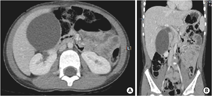
Fig. 4 Abdominal computed tomography (A, B) performed at 10 days after fever onset showed marked distended gallbladder (8.0×5.5 cm in size) without wall thickening or sludge, which is consistent with gallbladder hydrops.

Fig. 5 Abdominal ultrasonography (A) performed at 23 days after fever onset revealed a slow regression of the gallbladder hydrops (6.7×3.2 cm in size). Echocardiography (B) performed at 81 days after fever onset revealed a complete improvement in gallbladder hydrops and a normal gallbladder.
Reference
-
1. Taddio A, Pellegrin MC, Centenari C, Filippeschi IP, Ventura A, Maggiore G. Acute febrile cholestatic jaundice in children: keep in mind Kawasaki disease. J Pediatr Gastroenterol Nutr. 2012; 55:380–383.2. Kim DS. Kawasaki disease. Yonsei Med J. 2006; 47:759–772.
Article3. Eladawy M, Dominguez SR, Anderson MS, Glodé MP. Kawasaki disease and the pediatric gastroenterologist: a diagnostic challenge. J Pediatr Gastroenterol Nutr. 2013; 56:297–299.4. Yi DY, Kim JY, Choi EY, Choi JY, Yang HR. Hepatobiliary risk factors for clinical outcome of Kawasaki disease in children. BMC Pediatr. 2014; 14:51.
Article5. Chen CJ, Huang FC, Tiao MM, Huang YH, Lin LY, Yu HR, et al. Sonographic gallbladder abnormality is associated with intravenous immunoglobulin resistance in Kawasaki disease. ScientificWorldJournal. 2012; 2012:485758.
Article6. Newburger JW, Takahashi M, Gerber MA, Gewitz MH, Tani LY, Burns JC, et al. Diagnosis, treatment, and long-term management of Kawasaki disease: a statement for health professionals from the Committee on Rheumatic Fever, Endocarditis and Kawasaki Disease, Council on Cardiovascular Disease in the Young, American Heart Association. Circulation. 2004; 110:2747–2771.
Article7. Zulian F, Falcini F, Zancan L, Martini G, Secchieri S, Luzzatto C, et al. Acute surgical abdomen as presenting manifestation of Kawasaki disease. J Pediatr. 2003; 142:731–735.
Article8. Slovis TL, Hight DW, Philippart AI, Dubois RS. Sonography in the diagnosis and management of hydrops of the gallbladder in children with mucocutaneous lymph node syndrome. Pediatrics. 1980; 65:789–794.
Article9. Dinulos J, Mitchell DK, Egerton J, Pickering LK. Hydrops of the gallbladder associated with Epstein-Barr virus infection: a report of two cases and review of the literature. Pediatr Infect Dis J. 1994; 13:924–929.10. Yi DY, Chang EJ, Kim JY, Lee EH, Yang HR. Age, predisposing diseases, and ultrasonographic findings in determining clinical outcome of acute acalculous inflammatory gallbladder diseases in children. J Korean Med Sci. 2016; 31:1617–1623.
Article11. Suddleson EA, Reid B, Woolley MM, Takahashi M. Hydrops of the gallbladder associated with Kawasaki syndrome. J Pediatr Surg. 1987; 22:956–959.
Article12. Mathai SS, Kulkarni VB, Harsh P. Gall bladder hydrops - a rare initial presentation of Kawasaki disease. Indian J Pediatr. 2013; 80:616–617.
Article13. Egritas O, Nacar N, Hanioglu S, Soyer T, Tezic T. Early but prolonged gallbladder hydrops in a 7-month-old girl with Kawasaki syndrome: report of a case. Surg Today. 2007; 37:162–164.
Article14. Bosse KR, Lee GE, Treat JR, Small RM. A 7-year-old boy with abdominal pain, fever, and rash. Pediatr Ann. 2013; 42:275–277.
Article15. Kuijpers TW, Wiegman A, van Lier RA, Roos MT, Wertheim-van Dillen PM, Pinedo S, et al. Kawasaki disease: a maturational defect in immune responsiveness. J Infect Dis. 1999; 180:1869–1877.
Article
- Full Text Links
-
- Actions
-
Cited
- CITED
-
- Close
- Share
-
- Similar articles
-
- A Case of Kawasaki Disease Compicated by Hydrops of Gallbladder
- Mucocutaneous Lymph Node Syndrome: Report of one case complicated by gallbladder hydrops and diagnosed by ultrasound
- Sonography in the Diagnosis and Management of Hydrops of the Gallbladder in Childern with Mucocutaneous Lymph Node Syndrome
- Hydrops of the gallbladder in children
- A History for Experimental Animal Models of Endolymphatic Hydrops
