Ann Dermatol.
2016 Feb;28(1):119-120. 10.5021/ad.2016.28.1.119.
A Case of Acute-Onset, Painful Acral Granuloma Annulare
- Affiliations
-
- 1Department of Dermatology, Veterans Health Service Medical Center, Seoul, Korea. zooooz@hanmail.net
- KMID: 2429496
- DOI: http://doi.org/10.5021/ad.2016.28.1.119
Abstract
- No abstract available.
MeSH Terms
Figure
-
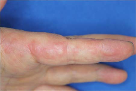
Fig. 1 Tender erythematous annular plaques on the lateral side of second and third finger.
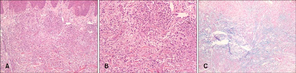
Fig. 2 (A) Palisade of histiocytes surrounding degenerated collagen and mucin in the dermis. A broad zone of mucinous degenerated collagen fibers with infiltrating lymphohistiocytic granuloma (H&E, ×100). (B) (H&E, ×400). (C) Alcian blue (pH 2.5) staining highlighted mucin in the center of the granuloma (×100).
Reference
-
1. Brey NV, Malone J, Callen JP. Acute-onset, painful acral granuloma annulare: a report of 4 cases and a discussion of the clinical and histologic spectrum of the disease. Arch Dermatol. 2006; 142:49–54.2. Stewart LR, George S, Hamacher KL, Hsu S. Granuloma annulare of the palms. Dermatol Online J. 2011; 17:7.
Article3. Gutte R, Kothari D, Khopkar U. Granuloma annulare on the palms: a clinicopathological study of seven cases. Indian J Dermatol Venereol Leprol. 2012; 78:468–474.
Article4. Hsu S, Lehner AC, Chang JR. Granuloma annulare localized to the palms. J Am Acad Dermatol. 1999; 41:287–288.
Article5. Na CH, Kim MS, Song SH, Shin BS. Solitary granuloma annulare: The first case of development on a healthy child's palm. Ann Dermatol. 2014; 26:113–114.
Article
- Full Text Links
-
- Actions
-
Cited
- CITED
-
- Close
- Share
-
- Similar articles
-
- Two Cases of Generalized Granuloma Annulare in Early Childhood
- Generalized Actinic Granuloma annulare with Impaired Glucose Tolerance Test
- Anetoderma due to Generalized Perforating Granuloma Annulare
- A Case of Generalized Granuloma Annulare
- A Case of Generalized Granuloma Annulare That Clinically Presented as Erythema Annulare Centrifugum
