Effects of prestretch on stress relaxation and permanent deformation of orthodontic synthetic elastomeric chains
- Affiliations
-
- 1Department of Orthodontics, College of Dentistry, Yonsei University, Seoul, Korea. yumichael@yuhs.ac
- 2Institute of Craniofacial Deformity, College of Dentistry, Yonsei University, Seoul, Korea.
- 3Department of Orthodontics, Gangnam Severance Dental Hospital, College of Dentistry, Yonsei University, Seoul, Korea.
- 4Department and Research Institute of Dental Biomaterials and Bioengineering, College of Dentistry, Yonsei University, Seoul, Korea.
- KMID: 2427776
- DOI: http://doi.org/10.4041/kjod.2018.48.6.384
Abstract
OBJECTIVE
This study was performed to investigate an appropriate degree of prestretch for orthodontic synthetic elastomeric chains focusing on time-dependent viscoelastic properties.
METHODS
Orthodontic synthetic elastomeric chains of two brands were prestretched to 50, 100, 150, and 200% of the original length in one and three cycles, and the hysteresis areas of the obtained stress-strain curves were determined. Acrylic plates were employed to maintain constant strain during the experiment. A total of 180 samples were classified into nine groups according to brand, and their stresses and permanent deformations were measured immediately after prestretch (0 hour), after 1 hour and 24 hours, and after 1, 2, 3, 4, 5, 6, 7, and 8 weeks. The relationship between stress relaxation and permanent deformation was investigated for various degrees of prestretch, and the estimated stress resulting from tooth movement was calculated.
RESULTS
The degree of prestretch and the stress relaxation ratio exhibited a strong negative correlation, whereas no correlation was found between the degree of prestretch and the average normalized permanent strain. The maximal estimated stress was observed when prestretch was performed in three cycles to 200% of the original length.
CONCLUSIONS
Although prestretch benefited residual stress, it did not exhibit negative effects such as permanent deformation. The maximal estimated stress was observed at the maximal prestretch, but the difference between prestretch and control groups decreased with time. In general, higher residual stresses were observed for product B than for product A, but this difference was not clinically significant.
Keyword
Figure
-
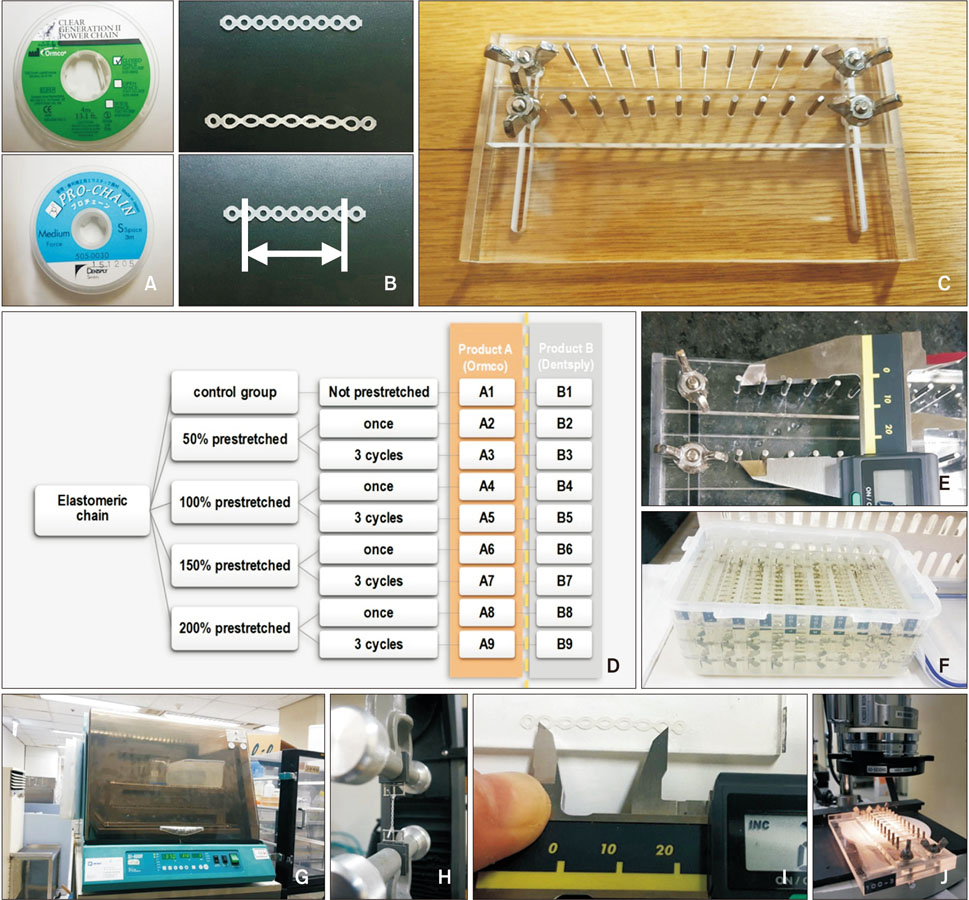
Figure 1 A, Orthodontic synthetic elastomeric chains used in the present study: product A (above) and product B (below). B, Photograph of control vs. permanent deformation (above), with the measured length indicated by an arrow (below). C, Fabricated acrylic plate. D, Classification table (product A: A1–A9; product B: B1–B9). E, Distance measurement for constant strain. F, Storage in artificial saliva. G, Storage in an incubator at 37℃. H, Measurement of stress using a universal testing machine. I, Distance measurement utilizing a digital Vernier caliper. J, Observation using the Micro Hi Scope system (Hirox, Tokyo, Japan).
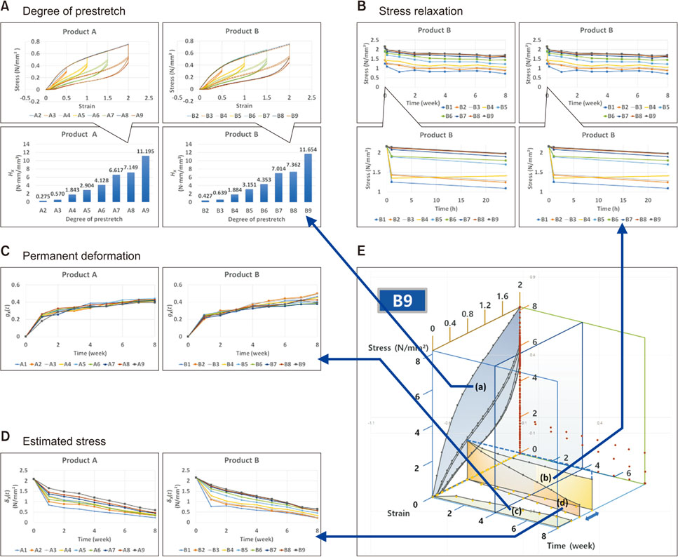
Figure 2 A, Plot of the hysteresis area enclosed by the stress-strain curve vs. the degree of prestretch. B, Stress relaxation curves measured for 8 weeks and enlarged curves recorded at times of up to 24 hours. C, Temporal dependence of normalized permanent strain (gx(t), Eq. 10). D, Temporal dependence of estimated stress (δx(t), Eq. 15) resulting from tooth movement (1 mm/4 weeks). E, Three-dimensional plot for group B9. Hysteresis representing the degree of prestretch is shown as the area of the stress-strain curve (a). A decrease in stress (b) and an increase in permanent deformation (c) were observed with increasing time. The reduction of estimated stress resulting from tooth movement (arrow) is shown in (d).
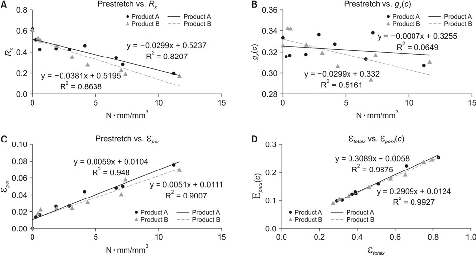
Figure 3 A, Scatter plot of the degree of prestretch (HX) vs. the stress relaxation ratio (RX). B, Scatter plot of HX vs. average normalized permanent strain (gX(c)). C, Scatter plot of HX vs. permanent deformation strain (εper) immediately after prestretch. D, Scatter plot of sustained total strain (εtotalx) vs. average permanent strain (εperx(c)).
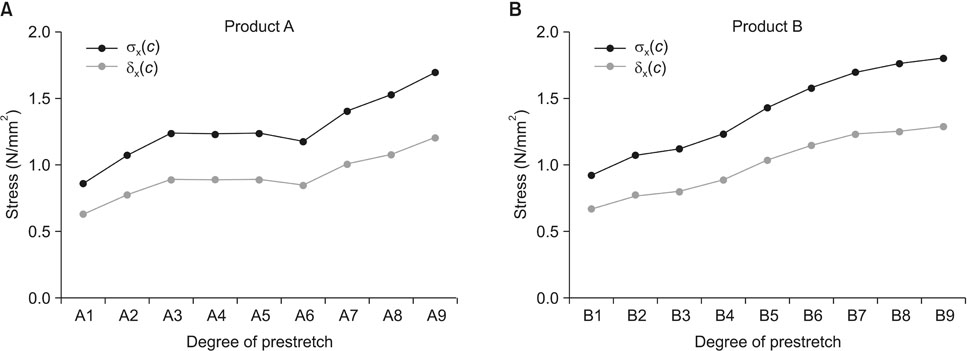
Figure 4 Average of recorded stress (σx(c)) and elastic restoring stress (δx(c)) of products A (A) and B (B) obtained after 8 weeks.
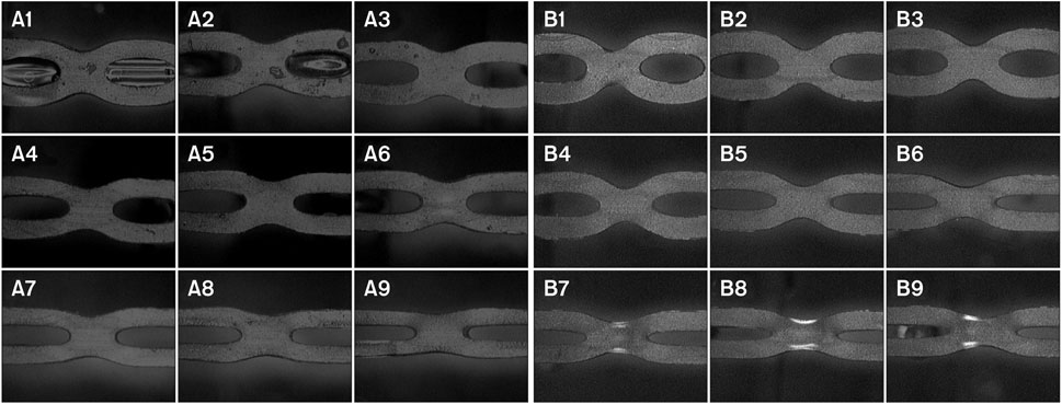
Figure 5 Images captured immediately after prestretch by the Micro Hi Scope system (Hirox, Tokyo, Japan; original magnification, ×50).
Reference
-
1. Halimi A, Benyahia H, Doukkali A, Azeroual MF, Zaoui F. A systematic review of force decay in orthodontic elastomeric power chains. Int Orthod. 2012; 10:223–240.
Article2. Andreasen GF, Bishara S. Comparison of alastik chains with elastics involved with intra-arch molar to molar forces. Angle Orthod. 1970; 40:151–158.3. Brantley WA, Salander S, Myers CL, Winders RV. Effects of prestretching on force degradation characteristics of plastic modules. Angle Orthod. 1979; 49:37–43.4. Wong AK. Orthodontic elastic materials. Angle Orthod. 1976; 46:196–205.5. Chang HF. Effects of instantaneous prestretching on force degradation characteristics of orthodontic plastic modules. Proc Natl Sci Counc Repub China B. 1987; 11:45–53.6. von Fraunhofer JA, Coffelt MT, Orbell GM. The effects of artificial saliva and topical fluoride treatments on the degradation of the elastic properties of orthodontic chains. Angle Orthod. 1992; 62:265–274.7. Stevenson JS, Kusy RP. Force application and decay characteristics of untreated and treated polyurethane elastomeric chains. Angle Orthod. 1994; 64:455–464.8. Kim KH, Chung CH, Choy K, Lee JS, Vanarsdall RL. Effects of prestretching on force degradation of synthetic elastomeric chains. Am J Orthod Dentofacial Orthop. 2005; 128:477–482.
Article9. Leung VWH, Darvell BW. Calcium phosphate system in saliva-like media. J Chem Soc-Faraday Trans. 1991; 87:1759–1764.
Article10. Yagura D, Baggio PE, Carreiro LS, Takahashi R. Deformation of elastomeric chains related to the amount and time of stretching. Dental Press J Orthod. 2013; 18:136–142.
Article11. De Genova DC, McInnes-Ledoux P, Weinberg R, Shaye R. Force degradation of orthodontic elastomeric chains--a product comparison study. Am J Orthod. 1985; 87:377–384.
Article12. Taloumis LJ, Smith TM, Hondrum SO, Lorton L. Force decay and deformation of orthodontic elastomeric ligatures. Am J Orthod Dentofacial Orthop. 1997; 111:1–11.
Article13. Storey EE, Smith R. Force in orthodontics and its relation to tooth movement. Aust J Dent. 1952; 56:11–18.14. Reitan K. Some factors determining the evaluation of forces in orthodontics. Am J Orthod Dentofacial Orthop. 1957; 43:32–45.
Article15. Lu TC, Wang WN, Tarng TH, Chen JW. Force decay of elastomeric chain--a serial study. Part II. Am J Orthod Dentofacial Orthop. 1993; 104:373–377.
Article16. Young J, Sandrik JL. The influence of preloading on stress relaxation of orthodontic elastic polymers. Angle Orthod. 1979; 49:104–109.17. Song HS, Kim SC. A study on the biomechanical properties of orthodontic rubber elastic materials. Korean J Orthod. 1991; 21:563–580.18. Kochenborger C, Silva DLd, Marchioro EM, Vargas DA, Hahn L. Assessment of force decay in orthodontic elastomeric chains: an in vitro study. Dent Press J Orthod. 2011; 16:93–99.19. Baty DL, Storie DJ, von Fraunhofer JA. Synthetic elastomeric chains: a literature review. Am J Orthod Dentofacial Orthop. 1994; 105:536–542.
Article20. Bishara SE, Andreasen GF. A comparison of time related forces between plastic alastiks and latex elastics. Angle Orthod. 1970; 40:319–328.21. Boester CH, Johnston LE. A clinical investigation of the concepts of differential and optimal force in canine retraction. Angle Orthod. 1974; 44:113–119.22. Lim HC, Lim SH. Comparison of physical characteristics of elastomeric chains. Oral Biol Res. 2011; 35:22–29.
Article23. Yee JA, Türk T, Elekdağ-Türk S, Cheng LL, Darendeliler MA. Rate of tooth movement under heavy and light continuous orthodontic forces. Am J Orthod Dentofacial Orthop. 2009; 136:150.e1–150.e9. discussion 150-1.
Article24. Hershey HG, Reynolds WG. The plastic module as an orthodontic tooth-moving mechanism. Am J Orthod. 1975; 67:554–562.
Article
- Full Text Links
-
- Actions
-
Cited
- CITED
-
- Close
- Share
-
- Similar articles
-
- Relaxation of orthodontic elastics, elastomeric modules and chains
- Elastic force degradation of synthetic elastomeric chain
- Physical properties of various brands of elastomeric chains
- Repetitive prestretching decreases hysteresis and abrupt force drops during the initial unloading phase of orthodontic elastomeric chains
- The second molars in orthodontics
