Magnifying Endoscopy for Esophageal Ectopic Sebaceous Glands
- Affiliations
-
- 1Department of Internal Medicine, Pusan National University School of Medicine, Busan, Korea. doc0224@pusan.ac.kr
- 2Department of Medicine and Pathology, Pusan National University School of Medicine, Busan, Korea.
- KMID: 2427724
- DOI: http://doi.org/10.5946/ce.2017.187
Abstract
- Ectopic sebaceous glands are found very rarely in the esophagus; heretofore, several cases have been reported. The sebaceous gland is originally a source of an endodermal origin; however, there have been controversies regarding whether the origin of the esophageal ectopic sebaceous gland is ectodermal or endodermal. Ectopic sebaceous glands of the esophagus usually do not cause symptoms; thus, they are often found incidentally on endoscopy for routine health screening. Endoscopic findings are characterized by single or multiple yellow patches or nodular lesions of various sizes, sometimes with small central openings. We report two cases of esophageal ectopic sebaceous glands found incidentally during endoscopy with magnifying endoscopic findings. The lesions were in the mid-esophagus and lower esophagus, respectively, and both endoscopic findings were similar as multiple yellowish patches or plaques. Magnifying endoscopy revealed the openings of the excretory ducts surrounded by circular microvessels in both cases.
Keyword
Figure
-

Fig. 1. (A) Upper endoscopy reveals multiple yellowish patches in the lower esophagus. (B) On magnifying endoscopy with narrow-band imaging, the excretory ducts of the sebaceous glands are slightly protruded and surrounded by circular microvessels (arrow). (C) Endoscopic biopsy reveals large and polygonal, clear cells with vacuolated cytoplasm within the squamous epithelium (hematoxylin and eosin, ×400).
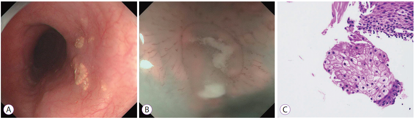
Fig. 2. (A) Upper endoscopy reveals several yellow plaques in the mid-esophagus. (B) Magnifying endoscopy with narrow-band imaging shows the excretory ducts of the sebaceous glands surrounded by circular microvessels. (C) Endoscopic biopsy reveals aggregates of polygonal cells with small central nuclei and abundant clear granular cytoplasm with foam-like fat droplets (hematoxylin and eosin, ×400).
Cited by 1 articles
-
Ectopic Sebaceous Gland in Esophagus Presenting as Subepithelial Tumor
Dong Han Yeom, Han Seung Ryu
Chonnam Med J. 2019;55(3):168-169. doi: 10.4068/cmj.2019.55.3.168.
Reference
-
1. Fordyce JA. A peculiar affection or the mucous membrane of the lips and oral cavity. Arch Dermatol. 1996; 132:1285.2. Bertoni G, Sassatelli R, Nigrisoli E, Conigliaro R, Bedogni G. Ectopic sebaceous glands in the esophagus: report of three new cases and review of the literature. Am J Gastroenterol. 1994; 89:1884–1887.3. De La Pava S, Pickren JW. Ectopic sebaceous glands in the esophagus. Arch Pathol. 1962; 73:397–399.4. Ramakrishnan T, Brinker JE. Ectopic sebaceous glands in the esophagus. Gastrointest Endosc. 1978; 24:293–294.
Article5. Bae JY, Chon CY, Kim H. Sebaceous glands in the esophagus. J Korean Med Sci. 1996; 11:271–274.
Article6. Kim SM, Im EH, Jung SH, et al. A case of ectopic sebaceous glands in the esophagus. Korean J Gastrointest Endosc. 2005; 31:320–322.
Article7. Shin JH, Jung JH, Choi HJ, Yoo J, Kang SJ, Lee KY. Ectopic sebaceous glands in the esophagus: a case report. Korean J Pathol. 2006; 40:448–451.8. Kim YS, Jin SY, Shim CS. Esophageal ectopic sebaceous glands. Clin Gastroenterol Hepatol. 2007; 5:A23.
Article9. Park HB, Cho HG, Kim YJ, et al. A case of ectopic sebaceous glands in the esophagus. Korean J Gastrointest Endosc. 2010; 41:155–158.10. Kim TH, Song JH, Kim TH, et al. A case of ectopic sebaceous glands in the esophagus. Korean J Helicobacter Up Gastrointest Res. 2012; 12:249–251.
Article11. Lee SH, Lee DJ, Kim KM, Kim KN, Kang JK. Ectopic sebaceous glands in the oesophagus: a case report and review of literature. Scott Med J. 2014; 59:e1–e5.
Article12. Chiu KW, Wu CK, Lu LS, Eng HL, Chiou SS. Diagnostic pitfall of sebaceous gland metaplasia of the esophagus. World J Clin Cases. 2014; 2:311–315.
Article13. Park A, Lee JH, Park A, et al. Prevalence rate and clinical characteristics of esophageal ectopic sebaceous glands in asymptomatic health screen examinees. Dis Esophagus. 2017; 30:1–5.
Article14. Rector LE. Aberrant mucosa in the esophagus in infants and in children. Arch Pathol. 1941; 31:285–294.15. Wang WP, Wang WS, Tsai YC. Multiple tiny ectopic sebaceous glands discovered throughout entire esophageal tract. Dig Dis Sci. 2009; 54:2754–2757.
Article16. Hoshika K, Inoue S, Mizuno M, Iida M, Shimizu M. Endoscopic detection of ectopic multiple minute sebaceous glands in the esophagus. Report of a case and review of the literature. Dig Dis Sci. 1995; 40:287–290.17. Wei IF, Chang CC, Fang CL, et al. Education and imaging. Gastrointestinal: ectopic sebaceous glands in the esophagus. J Gastroenterol Hepatol. 2008; 23:338.18. Bang CS, Kim YS, Baik GH, Han SH. Xanthoma of the esophagus. Clin Endosc. 2014; 47:358–361.
Article19. Nishisaki H, Yasutake K, Nakashima T, et al. Five cases with ectopic esophageal sebaceous glands. Dig Endosc. 1997; 9:207–212.
Article20. Fukuchi M, Tsukagoshi R, Sakurai S, et al. Ectopic sebaceous glands in the esophagus: endoscopic findings over three years. Case Rep Gastroenterol. 2012; 6:217–222.
Article
